Providing Remote Services and Support

By , March 24, 2020

The Center for the History of Medicine’s Holmes Hall reading room is currently closed due to COVID-19 precautions, but staff continue to provide remote support. Please contact chm@hms.harvard.edu for more information.

Incunabula volume, 1496

Conciliator differentiarum philosophorum et medicorum, 1496

Center staff provide the following services:

  • Schedule comprehensive consultations online (Zoom) or phone
  • Provide support for students, researchers and faculty during online office hours. (Please sign up in advance and a link will be provided.)
  • Assist in making arrangements for future visits and projects.

Online Resources:

Start your research remotely with the help of our collections’ online finding aids, and LibGuides. Other useful digital resources include:

  • OnView: the Center’s primary portal to digitized content and exhibits
  • Medical Heritage Library: online access to thousands of rare books, journals, pamphlets, and other items digitized as part of the Medical Heritage Library, Inc.’s consortium
  • Dataverse: Research data available from Center collections
  • Colonial North America: A Harvard Library project involving manuscript materials that relate to 17th- and 18th-century North America including items relating to science, medicine and health
  • Fredrick Stare papers: In collaboration with the University of California, San Francisco, the professional collection of Fredrick Stare, founder of the Department of Nutrition at the now T.H. Chan School of Public health, is digitally available.
Warren Anatomical Museum Gallery

Warren Anatomical Museum Gallery

For even more open access resources provided through Harvard Library, please check this evolving list.


Follow us for further information and updates on Twitter and Instagram @HarvardHistMed

The BackBlog: The Rise of Homeopathy

By , May 27, 2020

While some items in the backlog took lots of research to identify, this one did not. A card labeled “HOMEOPATHIC DRUG BOX” made the former contents of these bottles clear. 

Homeopathy is a medical practice based on the concept of “like cures like”. Homeopathic treatments are dilutions of a substance that would cause adverse symptoms in a healthy person but are believed to cure those symptoms in someone who is sick. For example, onion is used in remedies for seasonal allergies that result in itchy or sore eyes. Nux vomica, which comes from the strychnine tree, is used to treat nausea and hangovers. Most homeopathic preparations are so diluted that there are no longer any molecules of the original substance left. 

Photograph of a small chest containing small corked bottles. The chest is black. The lid is not attached and is sitting next to the part containing the bottles. A card that reads "HOMEOPATHIC DRUG BOX" is propped up against the chest.

Homeopathic Drug Box. From the Warren Anatomical Museum in the Center for the History of Medicine, Francis A. Countway Library of Medicine (WAM 22249)

The practice of homeopathy was founded in the late 18th century by the German physician Samuel Hahnemann (1755-1843). Hahnemann’s student, Hans Birch Gram, brought homeopathy to the United States in 1825. In the beginning, most practitioners of homeopathy—or “homeopaths”—were German immigrants. The first homeopathic medical school in the US was founded in Allentown, Pennsylvania in 1835.  The American Institute of Homeopathy was founded in 1844. Once these institutions were developed there was a rise in the popularity of homeopathy. More patients were interested in homeopathic treatments, and more Americans became practitioners. 

There were many reasons for this rise in popularity. One of the biggest was that homeopathy was considered safe compared to other medical treatments. Surgery often led to infection due to the lack of aseptic practices. The common treatments for diseases like cholera were unpleasant and ineffective. There were no active ingredients in most homeopathic remedies. Because of this, they could not cure an illness, but they also could not create negative side effects. Patients who were treated by homeopaths and recovered often reported a much better experience than their counterparts who had conventional medical treatments. 

Homeopathy also gained popularity because it was compared to another emerging medical practice: vaccination. Around the time that Hahnemann was creating his theory of homeopathy, Edward Jenner (1749-1823) was experimenting with giving people small doses of cowpox to prevent smallpox infection. Like homeopathy, vaccination involves exposing yourself to a small amount of something that would make you sick in a larger quantity. The two practices are different for many reasons, but this similarity is what people focused on. The success of vaccination likely led to the widespread use of homeopathy. 

Although it was widely used, homeopathy was not accepted by everyone in the medical field. One of the harshest critics was Oliver Wendell Holmes Sr. (1809-1894), who was dean of the Harvard Medical School from 1847-1853. In 1842 Holmes published a book titled Homœopathy and Its Kindred Delusions, based on two of his lectures. Holmes criticized the basis of homeopathy and stated his belief that the positive results patients saw were due to the placebo effect. He compared homeopathy to other ineffective medical treatments, like Perkins tractors and the royal touch—the belief that the laying on of hands by a monarch could cure a sick subject. 

Homeopathy lost popularity in the early 20th century as surgery became safer and new treatments became available but saw a resurgence in the 1970s. Homeopathic preparations are often sold in pharmacies alongside conventional treatments. While this case from the backlog and the tiny bottles inside of it might look different from the homeopathy that we see today, their contents were not dissimilar from what you might find on a shelf today. 

Oliver Wendell Holmes’ Friendship Cup

By , April 13, 2020

On August 29, 1889, former dean of Harvard Medical School Oliver Wendell Holmes (1809-1894) turned 80 years old. Annie Fields (1834-1915) and Sarah Orne Jewett (1849-1909), along with nine other women, presented Holmes with this silver loving cup at his birthday celebration. The cup entered the Harvard Medical Library collection in 1940 when Mrs. Richard Rule—the great-granddaughter of Holmes’ sister—presented it to the Department of Anatomy. A renewed focus was recently placed on the cup as part of an inventory project for this collection.  

Photograph of a stemless sliver loving cup with the engravings "Oliver Wendell Holmes" and "The Pledge of Friendship"

Oliver Wendell Holmes Friendship Cup. From the Harvard Medical Library in the Center for the History of Medicine, Francis A. Countway Library of Medicine (a001.098)

Holmes was the Dean of Harvard Medical School from 1847-1853 and was the Parkman Professor of Anatomy and Physiology until he retired in 1882. Holmes was a skilled physician who made great contributions to research in puerperal fever and was the first person to bring a microscope into an anatomy classroom in the United States. Along with his medical prowess, Holmes was also a prolific writer. He published both poetry and prose. He came up with the name for the magazine The Atlantic and contributed to it many times. These worlds often collided for Holmes: much of his writing revolved around the medical world, and he frequently gave recitations of his poetry at events for medical institutions. 


The 1889 loving cup—called the “Friendship Cup”—is engraved with a quote from Holmes’ poem “The Sentiment”. The engraving reads: 

The Pledge of Friendship 

Tis the heart’s current lends the cup its glow 

Whate’er the fountain whence the draught may flow 

Photograph of the bottom of the cup showing the eleven names engraved there: Helen C. Bell, Marianne Brimmer, Susan Cabot, Annie Fields, Alice G. Howe, Elizabeth Howes, Sarah O. Jewett, Mary G. Lodge, Minnie C. Pratt, Cora L. Shaw, and Sarah W. Whitman

The bottom of the loving cup

A loving cup is a shared drinking vessel and is usually used at weddings and other celebrations. In the 19th century, they were popular for trophies and commemorative gifts. Holmes was particularly enamored with the design of the cup. The names of the donors were on the bottom. In theory, the names could not be seen if the cup was full, so it would have to be emptied—and therefor shared—before they could be read. He highlighted this aspect of the cup in his poem, “To The Eleven Ladies Who Presented a Loving Cup to Me”, which begins: 

“Who gave this cup?” The secret thou wouldst steal 

 Its brimming flood forbids it to reveal: 

No mortal’s eye shall read it till he first 

Cool the red throat of thirst. 


If on the golden floor one draught remain, 

Trust me, thy careful search will be in vain; 

Not till the bowl is emptied shalt thou know 

The names enrolled below.

Originally, only twelve copies of this poem were printed: one for Holmes and one for each of the donors. He signed these copies by hand. Holmes later published the poem in his 1891 book Over the Teacupsa collection of poems and essays centered around fictional breakfast-table conversations. 

Scan of two pages that display the poem, "To The Eleven Ladies" by Oliver Wendell Holmes. Holmes' signature is underneath the printed poem.

One of Twelve Original Copies of “To The Eleven Ladies”, signed by Oliver Wendell Holmes. From the Boston Medical Library in the Center for the History of Medicine, Francis A. Countway Library of Medicine.

Not much is known about the donors or why they presented Holmes with this gift, but there are a few notable names on the bottom of the cup. Annie Fields was a writer who published collections of poetry and essays as well as biographies of notable literary figures. Her husband, James Fields, was a publisher. Together they ran literally salons out of their home, many of which were attended by Holmes. Annie Fields was also the sister of Zabdiel Boylston Adams, Jr., who was a surgeon and attended Harvard Medical School while Holmes was Dean. Sarah Orne Jewett was a friend and later companion of Annie Fields. She was also a writer and was best known for her work describing rural New England Life. Her father, Theodore Herman Jewett, was a doctor. Her experience accompanying him on his rounds as a child inspired her book A Country Doctor. 

It is easy to see why Fields and Jewett would have been drawn to Holmes, and although not much is known about the other donors, the Friendship Cup is a clear sign of admiration from all eleven women. Holmes’ poem indicates that the admiration was reciprocal. This reciprocity is part of what makes the Friendship Cup stand out in the collection: it doesn’t just reveal information about Holmes himself, but about his friendship with the donors. Although this loving cup was donated to the Harvard Medical Library as an artifact relating to Oliver Wendell Holmes, it sheds some light on the lives of eleven others as well. 

The BackBlog: J. W. Farlow’s Tools for Tonsillectomy

By , April 6, 2020

As I sorted through boxes of ear, nose, and throat instruments, I was excited to discover a clear collection forming. There were numerous instruments that had been carefully tagged with a description of the instrument and the name “Dr. J. W. Farlow”. This was particularly exciting because it wasn’t only a clear collection, but it all revolved around one particular subject. Before I knew anything about who J. W. Farlow was, I knew based on his collection that he was almost certainly a laryngologist.  

What was even more exciting was when I began looking at the names on the objects. Amongst objects with names like “Hajeck’s nasal punch” and “Roe’s septum forceps”, I found an object labeled “Farlow tonsil ecraseur”. J. W. Farlow wasn’t just a doctor, he was also an inventor.  

Photograph of a tonsil ecraseur. The object is made of metal and is shaped in a straight line with finger loops at the bottom and center of the object. There is no wire.

Farlow Tonsil Ecraseur. From the Warren Anatomical Museum in the Center for the History of Medicine, Francis A. Countway Library of Medicine (WAM 22220)

The instruments belonged to Dr. John Woodford Farlow (1853-1937), who was a laryngologist in Boston. At the time that Farlow was practicing, tonsillectomy—surgical removal of the tonsils—was a common procedure. In the early 20th century it was the most frequently practiced surgical procedure in the United States and would have made up a large portion of a laryngologist’s work. Because of this, there were many innovations in the tools used for these surgeries. Farlow made his mark with his tonsil forceps and the tonsil ecraseur (or tonsil snare) that is part of our collection.  

The tonsil ecraseur was an alternative to the tonsillotome or tonsil guillotine, allowing the physician to remove the tonsils in their entirety. It was often favored over other instruments because it removed the tonsils cleanly and because many believed—although it was disputed—that it reduced the chances of hemorrhage during surgery. Farlow’s ecraseur had a straight handle with a “cold wire”, meaning there was no heating element like some other devices. The wire was attached to a screw that could be rotated using the finger loops at the end of the handle. This allowed the physician using the device to carefully and gradually pull the wire, which gave Farlow’s design a distinct advantage over lever-based snares, which did not offer as much control. 

Farlow was associated with many hospitals in the Boston area throughout his career, including the Boston Dispensary, the Staniford Street Dispensary, the Free Dispensary for Diseases of Women, and the Boston City Hospital. He was on the Harvard Medical School faculty as a Clinical Instructor of Laryngology from 1892-1906. In 1902 he served as President of the American Laryngological Society. He was also very involved in the Boston Medical Library, which was the subject of most of his publications. Because of this, he is remembered more as a librarian than a laryngologist. His tonsil ecraseur and the rest of his collection are a reminder of his important contributions to his medical field.

How One Hospital Handled the 1918 Influenza Epidemic

By , April 2, 2020
Three doctors at the Peter Bent Brigham Hospital in Boston, MA outfitted for handling the influenza epidemic of 1918.

Doctors at the Peter Bent Brigham Hospital in Boston, MA outfitted for handling the influenza epidemic of 1918.

In 1918, no hospital, including the Peter Bent Brigham Hospital, a parent institution of the Brigham and Women’s Hospital, was spared the responsibility of caring for those afflicted by the worldwide influenza epidemic. The records of their battle with the mysterious illness endure in the BWH Archives held in the Center for the History of Medicine, Countway Library.

I say mysterious, because there was very little known about viruses or routes of contagion in the early years of the twentieth century. It would be more than a decade before the viral nature of influenza was uncovered and another quarter-century before the development of a vaccine or the widespread use of antibiotics for secondary bacterial infections. At the time, hospitals such as the Brigham could offer influenza patients supportive care (fluids, oxygen delivery, attention to heart, bowels, etc.). Also considered therapeutic—a good dose of fresh air.

Ventilation

The current Brigham and Women’s Hospital complex evolved on the site of the original Peter Bent Brigham Hospital. The Peter was designed in 1912 in the pavilion style, a hospital building system for which Florence Nightingale had been a strong advocate in the late Victorian era. The idea was that disease was probably spread in the “miasmic” air around the sick. This meant that patient wards were built as physically separated buildings designed for maximum cross-ventilation, with extra space between patients, and access to the outside. The wards were connected to each other and the main hospital building by an outdoor walkway.

The patient wards of the Peter Bent Brigham Hospital in 1918.

The patient wards of the Peter Bent Brigham Hospital in 1918. Note the outdoor beds for flu victims. (The ward pavilions were torn down in 1980.)

Flu patients in the Peter Bent Brigham Hospital recovering outdoors on the ward porch in 1918.

Flu patients in the Peter Bent Brigham Hospital recovering outdoors on the ward porch in 1918. Many archival photographs show patients recovering from illness outside in the early years of the hospital.

A New Warfront

When the influenza epidemic hit Boston, Dr. Henry A. Christian, the first Physician-in-Chief of the Peter Bent Brigham Hospital, was already dealing with a shortage of the experienced doctors and nurses who had taken leave to serve at military hospitals during the first World War—the armistice coming too late in the year to cause an appreciable change in staffing. Though filled to capacity and understaffed, the Brigham’s doctors, nurses, students, and volunteers worked selflessly to help the afflicted.

The best account of how the hospital managed the unexpected avalanche of influenza patients and the toll on his staff is from Dr. Christian’s own 1918 report:


The Influenza Epidemic

Henry A. ChristianIn the year 1918 no hospital event exceeded in importance and seriousness the epidemic of influenza, which burst upon Boston in September, and at the close of the year, was still prevalent, though to a lessened-degree. For a number of weeks practically the entire hospital was given over to the care of influenza cases. Every medical bed and half of the surgical beds were occupied by influenza cases. In the remaining beds were concentrated such surgical and medical cases as must needs remain in the hospital or be admitted as emergency cases. The Surgical Staff loaned to the Medical four of their house officers to care for influenza cases, and very generously the surgeons curtailed their work to a minimum. Without this help from the Surgical Staff we would have been unable to meet the needs of the situation.

The hospital cooperated with the Board of Health and took, in the main, cases selected by them, patients who could not be cared for at home, or those in almost dying condition that it was necessary to get out of their homes to ease the problem of home management of less seriously sick ones. Many died in a few hours after being brought to the hospital. The wards were filled with patients extremely ill with the pneumonia that accompanied influenza. More than half of our nursing and medical staffs themselves had influenza, but we managed to carry on without any outside help and without using probationers [newly admitted student nurses] or nurses’ aids in the immediate care of patients, feeling that for the less well trained the dangers of acquiring the disease from contact was greater.

From September 9 to December 31 we treated 557 cases of influenza. Of these about 60 per cent had clinically demonstrable pneumonia, a large percent being admitted to the hospital with pneumonia already developed. Of the influenza cases 153 died. A striking contrast to the general hospital cases were our nurses and doctors, who, put at once to bed and kept there, recovered with one exception. It seems certain that failure to go to bed and remain there was an important cause of the high mortality of the disease throughout the country. It had seemed wise to us to insist on a prolonged stay in bed after the temperature was normal. The very few relapses that we saw seemed amply to justify this course. No patients were hurried out of the hospital; all were urged to stay until we felt sure that their strength made the exertion of home life and getting there safe. In following up our patients since discharge we have found that results appear to show the wisdom of this course, for bad after-effects of the disease have been surprisingly few in our patients.

In handling influenza patients all who came in contact with them were gowned, capped, and masked, and care in washing hands was insisted on. Our present knowledge of influenza is too inadequate to make certain how far these precautions are necessary. At present they seem wise. Consideration of the infection among our nurses and doctors indicates that they acquired the disease not from patients in the wards but from contact with one another, or with maids, or with outside friends just developing the disease; in other words, the disease is most contagious during the incubation period. Only one non-influenza patient in the hospital acquired the disease in the hospital wards, and one patient acquired influenza in the operating room, probably from one of the doctors who assisted in operating on him as the doctor shortly thereafter developed influenza. Thus it would seem reasonably easy to maintain quarantine, even within an infected institution, when all visitors are excluded and other means of isolation are carried out. Such rigid quarantine probably is not possible in the community at large…

…Our nurses did most excellent work during the epidemic. The numerous cases of the disease among them made it necessary for the well ones to work with redoubled energy. Pupil nurses had to replace graduate head nurses as the latter fell ill, and so had thrust upon them much responsibility. Nurses could not but feel the grave danger they ran in handling a disease which evidently in some part of its course was extremely contagious and which was causing a high toll of deaths right under their eyes. There was no faltering; each did her duty and carried out her work efficiently. Particularly did the night nurses have a strenuous time, for in the silent hours of the night delirium was most active, and then it was that death claimed its largest numbers. The way our nurses met these demands upon them has caused the staff to feel great pride in them, and has been, I am sure, an occasion for much gratification to Miss Ivers and her associates who are immediately responsible for the training of the nurses. Personally it is a pleasure to me to voice here the feeling which all of the Medical Staff shares.

Leone N. Ivers, R.N., Acting Superintendent of Nurses, reported the situation for her nurses that same year:

On September 16, 1918, twelve nurses came down with influenza, and this epidemic was very serious during the balance of the year; the blackest day starting with forty-five nurses off duty. It was necessary for about one month to increase to a ten-hour day, with one afternoon off each week and four hours on Sunday. Enough cannot be said of the faithfulness and devotion of the nurses able to continue on duty during this period. It is with sincere regret that I am obliged to record the death of two of our pupil nurses. Marion Louise Winslow died from influenza at the New York Nursery and Child’s Hospital, New York City, October 12, 1918. She would have graduated January 26, 1919. Mabel Downton died from influenza at this hospital December 28, 1918. She was just starting on her senior year.

The full reports of all the administrators of the Peter Bent Brigham Hospital in 1918 can be read in the online version of the Peter Bent Brigham Hospital Annual Report for that year:
https://iiif.lib.harvard.edu/manifests/view/drs:427307276$1i

Nathan Cooley Keep and the Parkman-Webster Murder Trial

By , March 31, 2020

In 1846, Nathan Cooley Keep, a dentist, fashioned a set of false teeth for his patient George Parkman. A renewed focus was recently placed on the casts used to create these false teeth as part of an inventory project for the artifact collection of Harvard Medical Library. When he made these casts, Keep had no idea that a few years later, this work would lead to him testifying in a murder trial and giving the first piece of forensic dental evidence. 

Photograph of a plaster cast of inferior dentition

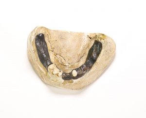
Inferior dental cast. From the Harvard Medical Library in the Center for the History of Medicine, Francis A. Countway Library of Medicine (a002.031)

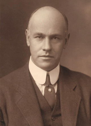
George Parkman (1790-1849) was a doctor from a wealthy family and a well-known part of the Boston elite. Parkman graduated from Harvard Medical College in 1813 and traveled to Europe to continue his studies. While Parkman remained a medical philanthropist throughout his life, he turned his career to real estate and developed his fortune even further. Parkman had bought up so much land in Boston that he donated some of it to the Medical School at Harvard so that they could relocate from Cambridge to Boston. Parkman also began to lend money and was known for his long walks around the city to collect his debts as he was too thrifty to buy a horse. He was on one of these walks on November 23, 1849, when he was seen entering Harvard Medical College to speak with John Webster.  

John White Webster (1793-1850) was a lecturer of chemistry and geology at Harvard Medical College. Webster was also a part of Boston high society, but his salary could not cover his expenses and his family was forced to give up their Cambridge mansion. Webster began borrowing money from a number of friends in order to deal with his financial difficulties. One of these friends was George Parkman. Webster began borrowing from Parkman in 1842 and continued to borrow large sums without paying Parkman back. By 1849 he owed Parkman so much money that he took out a loan from another friend to repay him but had used the same mineral cabinet as collateral that he had used with Parkman. The two agreed to a meeting at the Medical College on November 23, 1849, in order to come to an agreement regarding Webster’s debt. Parkman was seen entering the building at 1:45.  

Later that day the janitor, Ephriam Littlefield, was surprised to find that Webster’s laboratory was locked from the inside. He could hear water running inside, although Webster was not there. Littlefield’s suspicions began to grow when news spread that Parkman, who he had seen entering the building on the 23rd, was reported missing. Prior to Parkman’s visit, Webster had asked Littlefield a number of questions regarding the dissection vault. A few days after the meeting Webster had asked Littlefield if he had seen Parkman and appeared agitated when Littlefield said he had. Webster had also given him a Thanksgiving turkey, which seemed strange to Littlefield as Webster had never given him a gift before.  

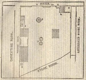
Floor plan of Webster’s laboratory from Trial of Professor John W. Webster for the Murder of Doctor George Parkman

Littlefield began to watch Webster closely. On November 28, 1849, he spied on Webster through the space underneath his door and watched Webster go back and forth between the furnace and the fuel closet several times. When Webster left, Littlefield broke into the laboratory through a window. He noticed that the kindling was nearly gone and there were strange spots of what looked like acid around the room. Over the Thanksgiving break, Littlefield began to excavate the wall underneath Webster’s private privy. After two days of digging, he uncovered a foul stench and what looked like a human pelvis. He called the police, who began a search. Eventually, a human pelvis, right thigh, and left calf were found in the privy. A jawbone and teeth were found in the furnace. John White Webster was arrested and put on trial for the murder of George Parkman.  

Because Parkman was such a well-known figure, the public was already obsessed with the case by the time of Webster’s arrest. When they learned the details of the charges against Webster, the case became even more salacious. Not only had Parkman been killed by another doctor, but he had also been dismembered and the pieces of his body had been hidden in the medical school. Newspaper headlines all revolved around Parkman and Webster, and tickets to the trials were so popular that spectators were being allowed in for a short time and then ushered out to make room for newcomers. It is estimated that thousands of people witnessed at least part of the trial.  

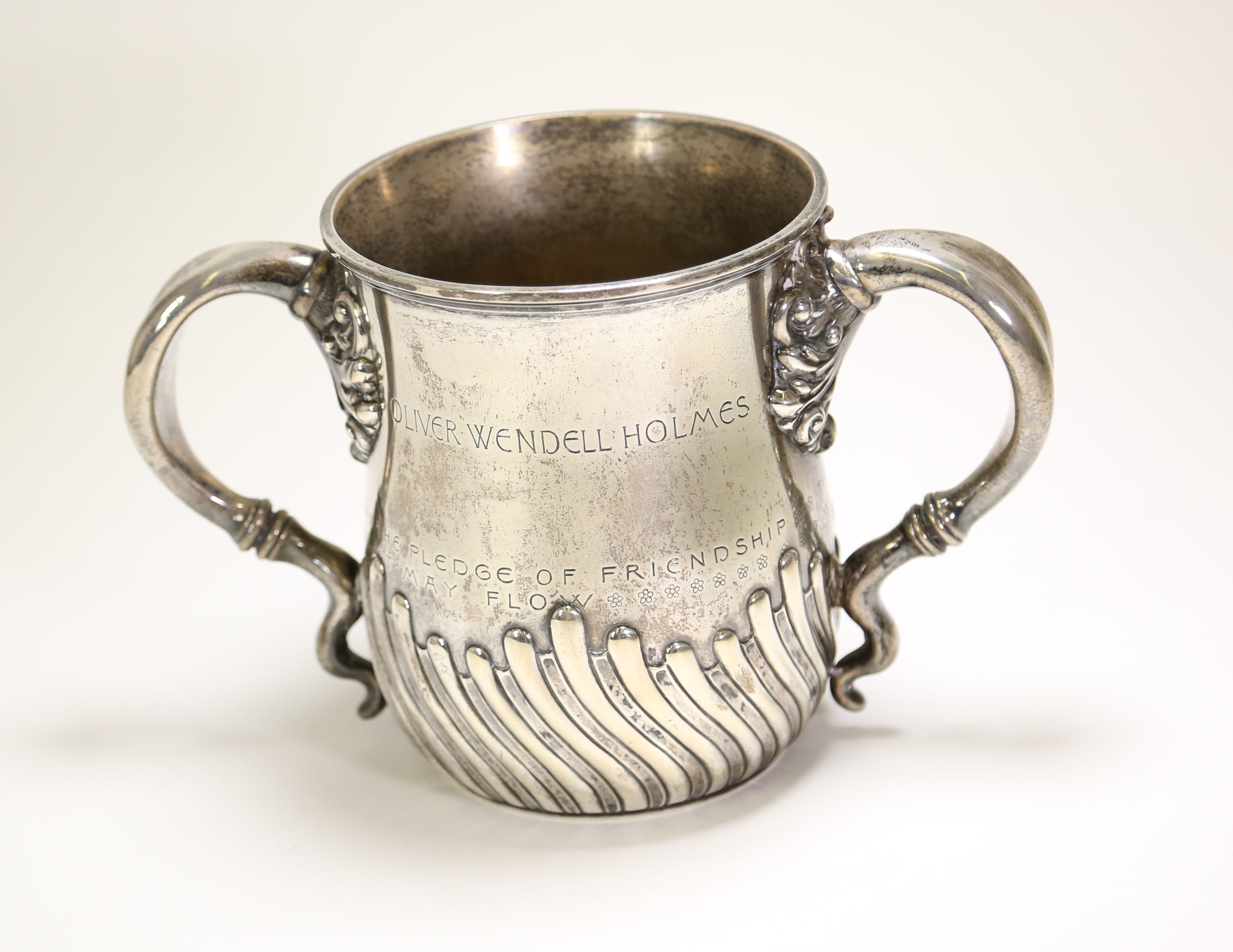
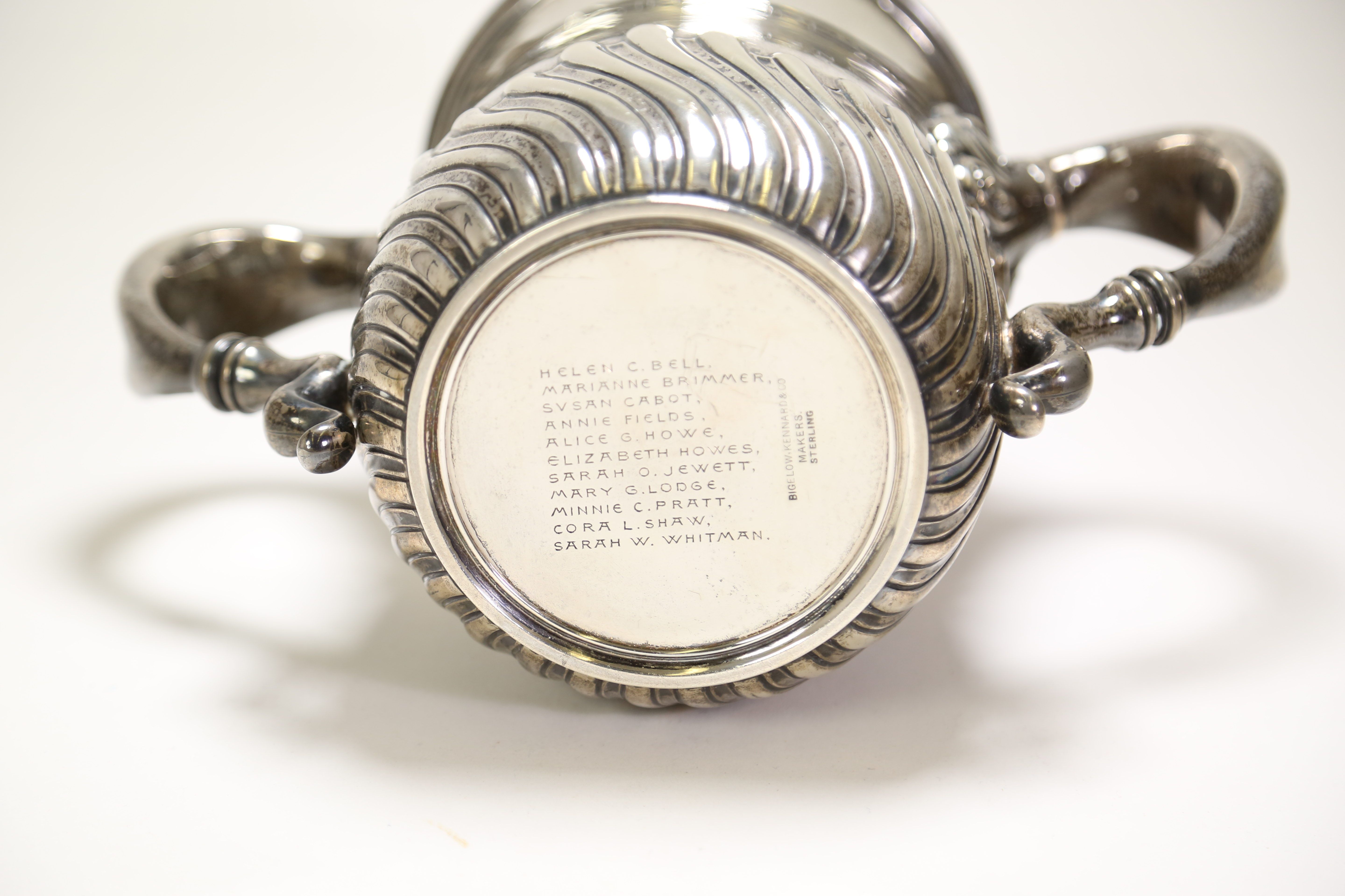
Because of the state in which the remains were found, the prosecution had to prove that the body was, in fact, that of George Parkman and that his death was a result of homicide. Multiple witnesses were called to the stand to confirm the identity of the body and the manner of death. Many of these people were Webster’s colleagues from the Medical College, including Jeffries Wyman (1814-1874), who testified that the remains matched the height and general size of Parkman, and Oliver Wendell Holmes (1809-1894), who testified that a small wound on the chest could have been a fatal stab wound. Ephriam Littlefield also testified, describing the events of the days leading up to and following Parkman’s disappearance.  

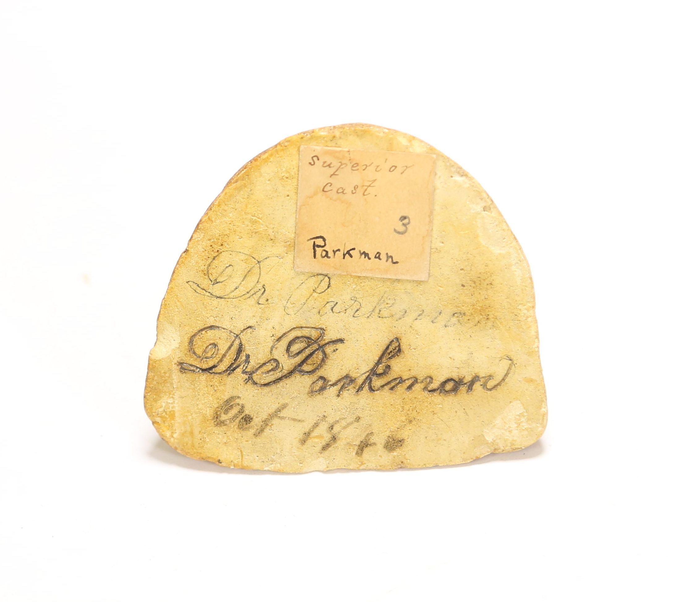
One of the most important pieces of testimony came from Parkman’s dentist, Nathan Cooley Keep (1800-1875). Keep, who later became the founding dean of the Harvard School of Dental Medicine, was a prominent dentist with high aspirations to unite dental and medical education at the time of the trial. He testified that the jawbone found in the furnace contained false teeth that he had made specifically for Parkman. Keep then showed the jury that the jawbone from the furnace fit perfectly into the plaster impression he had created of Parkman’s jaw in 1846 when he was fitting Parkman for the false teeth. Keep also showed them that loose teeth found in the furnace fit into the plates he had created. This convinced the jury that the body found in Webster’s laboratory was indeed George Parkman.  

Photograph of the underside of a dental plaster cast. "Dr. Parkman" is carved into the cast and "Oct 1846" is written on it.

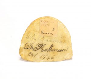
Bottom of superior dental cast showing Dr. Parkman’s name. From the Harvard Harvard Medical Library in the Center for the History of Medicine, Francis A. Countway Library of Medicine (a002.031)

John White Webster was found guilty of the murder of George Parkman on March 30, 1850, and was sentenced to be hanged. Although some still doubt his guilt, Webster confessed to the murder in June of 1850 and was executed on August 30, 1850.  

Although the case was famous at the time for its association with Boston high society, there is another reason that the trial of John Webster has lived on. This was the first time that dental evidence was accepted as part of a murder trial. These plaster casts were a vital part of the prosecution’s case, as they were the only piece of evidence or testimony that could confirm without a doubt that the body Littlefield had found was George Parkman’s. Over a century and a half after Keep introduced these plaster casts into evidence, dental evidence has become a key part of forensic science. Dental records are used to confirm identity today in much the same way that Keep used his plaster casts. The field has expanded over time, and when matching dental records cannot be found, teeth can be used to estimate identifying traits such as age and ancestry. George Parkman was the first person to be identified through forensic dentistry, but he was far from the last. 

The Parkman-Webster case was not only a media sensation. It was a landmark case in the history of forensic medicine. 

Now Accepting Applications for the 2020-2021 Women in Medicine Legacy Foundation Research Fellowship

By , March 24, 2020

The Women in Medicine Legacy Foundation Research Fellowship

Application deadline is May 31, 2020

Details

The Women in Medicine Legacy Foundation is pleased to provide one $5,000 grant to support travel, lodging, and incidental expenses for a flexible research period between July 1, 2019 and June 30, 2020. Foundation Fellowships are offered for research related to the history of women to be conducted at the Center for the History of Medicine at the Francis A. Countway Library of Medicine. Preference will be given to:

  • projects that engage specifically with the history of women physicians, other health workers, or medical scientists; proposals on the history of women’s health issues will also be considered
  • those who are using the Center’s Women in Medicine collections; however, research on the topic of women in medicine using other material from the Countway Library will be considered
  • applicants who live beyond commuting distance of the Countway; however, all are encouraged to apply, including graduate students

In return, the Foundation requests a one page report on the Fellow’s research experience, a copy of the final product (with the ability to post excerpts from the paper/project), and a photo and bio of the Fellow for web and newsletter announcements. The Fellow will also be asked to present a lecture at the Countway Library.

Women in Medicine Legacy Foundation logo

Women in Medicine Legacy Foundation logo

Application Requirements

Applicants should submit a proposal (no more than five pages) outlining the subject and objectives of the research project, length of residence, historical materials to be used, and a project budget (including travel, lodging, and research expenses), along with a curriculum vitae and two letters of recommendations by May 31, 2020. The fellowship proposal should demonstrate that the Countway Library has resources central to the research topic.

Applications and supporting documentation should be emailed to the Center for History of Medicine via chm@hms.harvard.edu. Please include “Women in Medicine Legacy Foundation Research Fellowship” in the subject line. Questions may be directed to chm@hms.harvard.edu or (617) 432-7702.


Partnering Organizations

The Women in Medicine Legacy Foundation, formerly the Foundation for the History of Women in Medicine, was founded with the strong belief that understanding our history plays a powerful role in shaping our future. The resolute stand women took to establish their place in these fields propels our vision forward. We serve as stewards to the stories from the past, and take pride in sharing them with the women of today. Our mission is to preserve and promote the history of women in medicine and the medical sciences, and we look forward to connecting you to our collective legacy that will empower our future.

The Archives for Diversity and Inclusion, formerly the Archives for Women in Medicine, is a program of the Countway Library’s Center for the History of Medicine at Harvard Medical School. The program’s goal is to ensure the Center’s collections reflect the diversity of the Harvard Medical School community by acquiring the research, teaching, and professional records of underrepresented faculty, including women. Learn more about collections open to research on our Women in Medicine Collections page.

Established in 1960 as a result of an alliance between the Boston Medical Library and the Harvard Medical Library, the Francis A. Countway Library of Medicine is the largest academic medical library in the United States. The Countway Library maintains a collection of approximately 700,000 volumes. The Center for the History of Medicine’s collection of archives and manuscripts, numbering between 15-20 million items, is the largest collection of its kind in the United States. Collections include the personal and professional records of physicians from the medieval and Renaissance periods through the twentieth century, including the professional papers of many renowned Harvard faculty members as well as physicians and scientists from New England and around the country.

The 2019-2020 Foundation for the History of Women in Medicine Research Fellow is Heather Munro Prescott, Ph.D. Previous fellows include Carla Bittel, Maria Daxenbichler, Jordan Katz, Kate Grauvogel, Louella McCarthy, Rebecca Kluchin, Ciara Breathnach, Carrie Adkins, and Hilary Aquino.


The BackBlog: Color Perception and Cards of Wool

By , March 19, 2020

This object came up in one of the first boxes we took off the shelf for our backlog project, while a group of Center staff was still trying to figure out exactly what our sorting process was going to be like. When we first opened the box and saw all of the yarncovered cards we were confusedThis looked more like a crafting set than a medical device. We were even a bit concerned that this box might have been donated to the museum in order to display a set of toxic dyes. But when we saw the name “B. Joy Jeffries” on the stationary in the box, we knew that it must be some sort of color blindness test. 

Photograph of a wooden box containing 24 wooden cards. Each card is wrapped in a different color of yarn with varying striped patterns.

Donders’ test for color blindness. From the Warren Anatomical Museum in the Center for the History of Medicine, Francis A. Countway Library of Medicine (WAM 22251)

Benjamin Joy (B. Joy) Jeffries (1833-1915) was a 19th century ophthalmologist. His work focused primarily on the causes and identification of color blindness. He even wrote a book titled: Color Blindness: Its Dangers and Its Detection. His writing on the subject is extensive and passionate. At the time that Jeffries was writing, color blindness was not commonly identified. It was a seriouhazard for railroad workers and people in similar occupations. A misinterpreted signal due to not seeing the right colors could result in a dangerous or even deadly situation. Jeffries’ work on the subject and advocacy for testing resulted in a much deeper understanding of color blindness and a safer railroad system. 

Identifying this particular test, however, was a bit of a challenge. It was labeled as “Holmgren’s Worsteds” in our accession record and on a label accompanying the objectHolmgren’s method of identification was Jeffries’ preferred method, and he wrote about it extensively. But while that test uses the same type of yarn, it involves matching small yarn bundles of the same color. Jeffries’ description made it clear that the object we found was not Holmgren’s test. 

Photograph of a piece of stationery with handwritten notes describing Donders' test for colorblindness. B. Joy Jeffries' name is imprinted at the top of the paper.

Description of test written on B. Joy Jeffries’ stationery, found with WAM 22251

Based on the methods listed in Jeffries’ book, this is most likely Donders’ test. This test was developed in 1879 by the ophthalmologist Franciscus Cornelis (F. C.) Donders (1818-1889). Jeffries describes Donders’ test as being made up of a set of wooden cards with different colors of wool wrapped around them. On some of the cards, a second color—one that a person who was color blind would not be able to differentiate from the firstwas wrapped over the first, and the subjects were asked to identify which cards had multiple colors. Like Holmgren’s test, Donders’ test involved a fairly simple procedure that wouldn’t have needed the complicated equipment like colored lights and spinning disks that some other methods required.  

Although he preferred Holmgren’s method, it is not surprising to find another type of testing amongst Jeffries’ collection. It is clear from his book that Jeffries tried every method for testing colorblindness that was available to him. We do have a few other color blindness tests in the museum, but as far as we know, this is the only one that belonged to B. Joy Jeffries. This simple wooden box with different colors of yarn—an object that I had originally thought looked like crafting supplies—turned out to be a fundamental piece in the history of color blindness research.

Center Reading Room Closed Until Further Notice

By , March 16, 2020

Due to coronavirus (COVID-19), starting Tuesday, March 17 and until further notice, the Center reading room is CLOSED to all researchers. We are able to provide limited online reference service.

Please contact us to schedule a remote consultation or ask a reference question at chm@hms.harvard.edu.


Panorama Theme by Themocracy