The BackBlog: The Rise of Homeopathy

By , May 27, 2020

While some items in the backlog took lots of research to identify, this one did not. A card labeled “HOMEOPATHIC DRUG BOX” made the former contents of these bottles clear. 

Homeopathy is a medical practice based on the concept of “like cures like”. Homeopathic treatments are dilutions of a substance that would cause adverse symptoms in a healthy person but are believed to cure those symptoms in someone who is sick. For example, onion is used in remedies for seasonal allergies that result in itchy or sore eyes. Nux vomica, which comes from the strychnine tree, is used to treat nausea and hangovers. Most homeopathic preparations are so diluted that there are no longer any molecules of the original substance left. 

Photograph of a small chest containing small corked bottles. The chest is black. The lid is not attached and is sitting next to the part containing the bottles. A card that reads "HOMEOPATHIC DRUG BOX" is propped up against the chest.

Homeopathic Drug Box. From the Warren Anatomical Museum in the Center for the History of Medicine, Francis A. Countway Library of Medicine (WAM 22249)

The practice of homeopathy was founded in the late 18th century by the German physician Samuel Hahnemann (1755-1843). Hahnemann’s student, Hans Birch Gram, brought homeopathy to the United States in 1825. In the beginning, most practitioners of homeopathy—or “homeopaths”—were German immigrants. The first homeopathic medical school in the US was founded in Allentown, Pennsylvania in 1835.  The American Institute of Homeopathy was founded in 1844. Once these institutions were developed there was a rise in the popularity of homeopathy. More patients were interested in homeopathic treatments, and more Americans became practitioners. 

There were many reasons for this rise in popularity. One of the biggest was that homeopathy was considered safe compared to other medical treatments. Surgery often led to infection due to the lack of aseptic practices. The common treatments for diseases like cholera were unpleasant and ineffective. There were no active ingredients in most homeopathic remedies. Because of this, they could not cure an illness, but they also could not create negative side effects. Patients who were treated by homeopaths and recovered often reported a much better experience than their counterparts who had conventional medical treatments. 

Homeopathy also gained popularity because it was compared to another emerging medical practice: vaccination. Around the time that Hahnemann was creating his theory of homeopathy, Edward Jenner (1749-1823) was experimenting with giving people small doses of cowpox to prevent smallpox infection. Like homeopathy, vaccination involves exposing yourself to a small amount of something that would make you sick in a larger quantity. The two practices are different for many reasons, but this similarity is what people focused on. The success of vaccination likely led to the widespread use of homeopathy. 

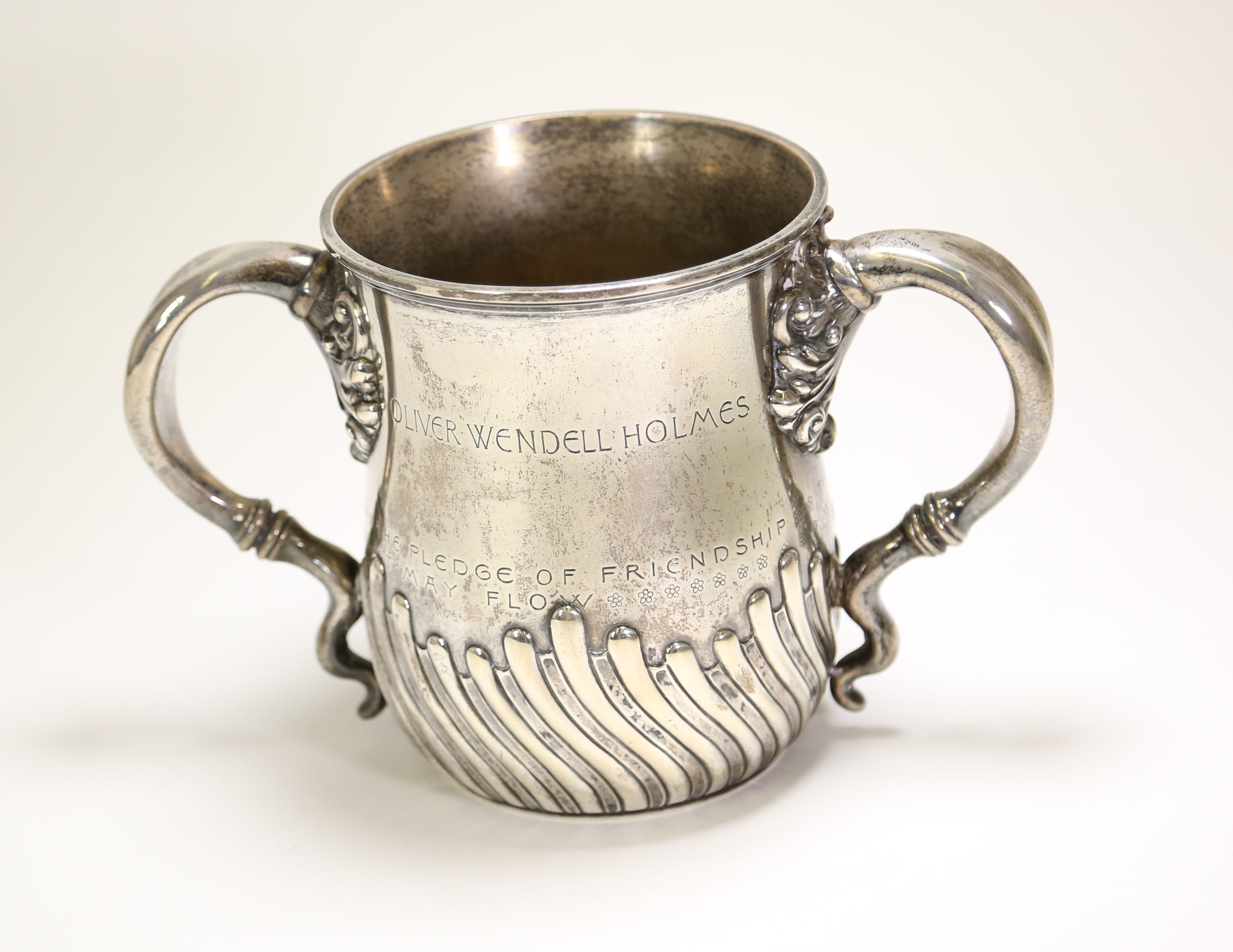
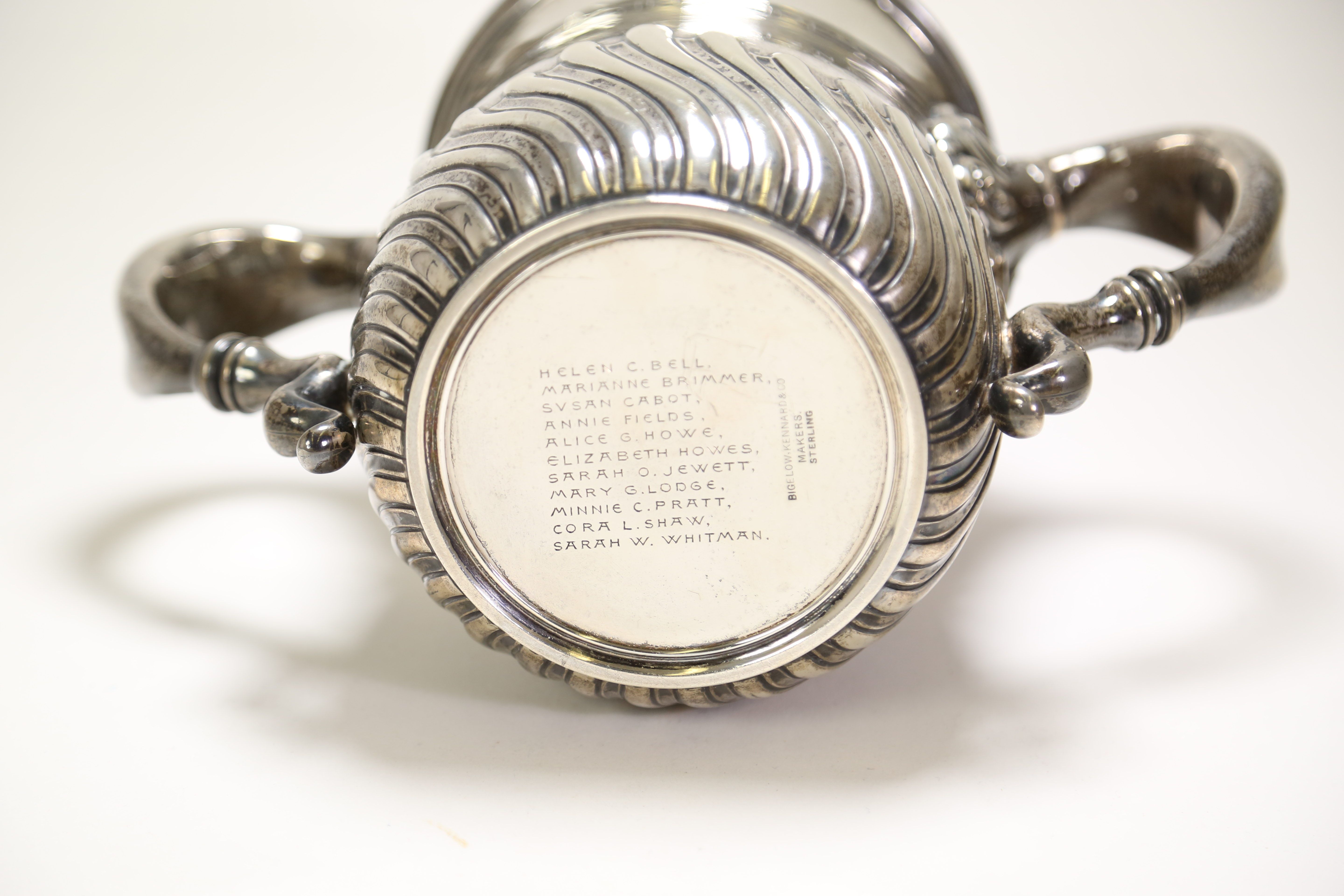
Although it was widely used, homeopathy was not accepted by everyone in the medical field. One of the harshest critics was Oliver Wendell Holmes Sr. (1809-1894), who was dean of the Harvard Medical School from 1847-1853. In 1842 Holmes published a book titled Homœopathy and Its Kindred Delusions, based on two of his lectures. Holmes criticized the basis of homeopathy and stated his belief that the positive results patients saw were due to the placebo effect. He compared homeopathy to other ineffective medical treatments, like Perkins tractors and the royal touch—the belief that the laying on of hands by a monarch could cure a sick subject. 

Homeopathy lost popularity in the early 20th century as surgery became safer and new treatments became available but saw a resurgence in the 1970s. Homeopathic preparations are often sold in pharmacies alongside conventional treatments. While this case from the backlog and the tiny bottles inside of it might look different from the homeopathy that we see today, their contents were not dissimilar from what you might find on a shelf today. 

Oliver Wendell Holmes’ Friendship Cup

By , April 13, 2020

On August 29, 1889, former dean of Harvard Medical School Oliver Wendell Holmes (1809-1894) turned 80 years old. Annie Fields (1834-1915) and Sarah Orne Jewett (1849-1909), along with nine other women, presented Holmes with this silver loving cup at his birthday celebration. The cup entered the Harvard Medical Library collection in 1940 when Mrs. Richard Rule—the great-granddaughter of Holmes’ sister—presented it to the Department of Anatomy. A renewed focus was recently placed on the cup as part of an inventory project for this collection.  

Photograph of a stemless sliver loving cup with the engravings "Oliver Wendell Holmes" and "The Pledge of Friendship"

Oliver Wendell Holmes Friendship Cup. From the Harvard Medical Library in the Center for the History of Medicine, Francis A. Countway Library of Medicine (a001.098)

Holmes was the Dean of Harvard Medical School from 1847-1853 and was the Parkman Professor of Anatomy and Physiology until he retired in 1882. Holmes was a skilled physician who made great contributions to research in puerperal fever and was the first person to bring a microscope into an anatomy classroom in the United States. Along with his medical prowess, Holmes was also a prolific writer. He published both poetry and prose. He came up with the name for the magazine The Atlantic and contributed to it many times. These worlds often collided for Holmes: much of his writing revolved around the medical world, and he frequently gave recitations of his poetry at events for medical institutions. 


The 1889 loving cup—called the “Friendship Cup”—is engraved with a quote from Holmes’ poem “The Sentiment”. The engraving reads: 

The Pledge of Friendship 

Tis the heart’s current lends the cup its glow 

Whate’er the fountain whence the draught may flow 

Photograph of the bottom of the cup showing the eleven names engraved there: Helen C. Bell, Marianne Brimmer, Susan Cabot, Annie Fields, Alice G. Howe, Elizabeth Howes, Sarah O. Jewett, Mary G. Lodge, Minnie C. Pratt, Cora L. Shaw, and Sarah W. Whitman

The bottom of the loving cup

A loving cup is a shared drinking vessel and is usually used at weddings and other celebrations. In the 19th century, they were popular for trophies and commemorative gifts. Holmes was particularly enamored with the design of the cup. The names of the donors were on the bottom. In theory, the names could not be seen if the cup was full, so it would have to be emptied—and therefor shared—before they could be read. He highlighted this aspect of the cup in his poem, “To The Eleven Ladies Who Presented a Loving Cup to Me”, which begins: 

“Who gave this cup?” The secret thou wouldst steal 

 Its brimming flood forbids it to reveal: 

No mortal’s eye shall read it till he first 

Cool the red throat of thirst. 


If on the golden floor one draught remain, 

Trust me, thy careful search will be in vain; 

Not till the bowl is emptied shalt thou know 

The names enrolled below.

Originally, only twelve copies of this poem were printed: one for Holmes and one for each of the donors. He signed these copies by hand. Holmes later published the poem in his 1891 book Over the Teacupsa collection of poems and essays centered around fictional breakfast-table conversations. 

Scan of two pages that display the poem, "To The Eleven Ladies" by Oliver Wendell Holmes. Holmes' signature is underneath the printed poem.

One of Twelve Original Copies of “To The Eleven Ladies”, signed by Oliver Wendell Holmes. From the Boston Medical Library in the Center for the History of Medicine, Francis A. Countway Library of Medicine.

Not much is known about the donors or why they presented Holmes with this gift, but there are a few notable names on the bottom of the cup. Annie Fields was a writer who published collections of poetry and essays as well as biographies of notable literary figures. Her husband, James Fields, was a publisher. Together they ran literally salons out of their home, many of which were attended by Holmes. Annie Fields was also the sister of Zabdiel Boylston Adams, Jr., who was a surgeon and attended Harvard Medical School while Holmes was Dean. Sarah Orne Jewett was a friend and later companion of Annie Fields. She was also a writer and was best known for her work describing rural New England Life. Her father, Theodore Herman Jewett, was a doctor. Her experience accompanying him on his rounds as a child inspired her book A Country Doctor. 

It is easy to see why Fields and Jewett would have been drawn to Holmes, and although not much is known about the other donors, the Friendship Cup is a clear sign of admiration from all eleven women. Holmes’ poem indicates that the admiration was reciprocal. This reciprocity is part of what makes the Friendship Cup stand out in the collection: it doesn’t just reveal information about Holmes himself, but about his friendship with the donors. Although this loving cup was donated to the Harvard Medical Library as an artifact relating to Oliver Wendell Holmes, it sheds some light on the lives of eleven others as well. 

The BackBlog: J. W. Farlow’s Tools for Tonsillectomy

By , April 6, 2020

As I sorted through boxes of ear, nose, and throat instruments, I was excited to discover a clear collection forming. There were numerous instruments that had been carefully tagged with a description of the instrument and the name “Dr. J. W. Farlow”. This was particularly exciting because it wasn’t only a clear collection, but it all revolved around one particular subject. Before I knew anything about who J. W. Farlow was, I knew based on his collection that he was almost certainly a laryngologist.  

What was even more exciting was when I began looking at the names on the objects. Amongst objects with names like “Hajeck’s nasal punch” and “Roe’s septum forceps”, I found an object labeled “Farlow tonsil ecraseur”. J. W. Farlow wasn’t just a doctor, he was also an inventor.  

Photograph of a tonsil ecraseur. The object is made of metal and is shaped in a straight line with finger loops at the bottom and center of the object. There is no wire.

Farlow Tonsil Ecraseur. From the Warren Anatomical Museum in the Center for the History of Medicine, Francis A. Countway Library of Medicine (WAM 22220)

The instruments belonged to Dr. John Woodford Farlow (1853-1937), who was a laryngologist in Boston. At the time that Farlow was practicing, tonsillectomy—surgical removal of the tonsils—was a common procedure. In the early 20th century it was the most frequently practiced surgical procedure in the United States and would have made up a large portion of a laryngologist’s work. Because of this, there were many innovations in the tools used for these surgeries. Farlow made his mark with his tonsil forceps and the tonsil ecraseur (or tonsil snare) that is part of our collection.  

The tonsil ecraseur was an alternative to the tonsillotome or tonsil guillotine, allowing the physician to remove the tonsils in their entirety. It was often favored over other instruments because it removed the tonsils cleanly and because many believed—although it was disputed—that it reduced the chances of hemorrhage during surgery. Farlow’s ecraseur had a straight handle with a “cold wire”, meaning there was no heating element like some other devices. The wire was attached to a screw that could be rotated using the finger loops at the end of the handle. This allowed the physician using the device to carefully and gradually pull the wire, which gave Farlow’s design a distinct advantage over lever-based snares, which did not offer as much control. 

Farlow was associated with many hospitals in the Boston area throughout his career, including the Boston Dispensary, the Staniford Street Dispensary, the Free Dispensary for Diseases of Women, and the Boston City Hospital. He was on the Harvard Medical School faculty as a Clinical Instructor of Laryngology from 1892-1906. In 1902 he served as President of the American Laryngological Society. He was also very involved in the Boston Medical Library, which was the subject of most of his publications. Because of this, he is remembered more as a librarian than a laryngologist. His tonsil ecraseur and the rest of his collection are a reminder of his important contributions to his medical field.

Nathan Cooley Keep and the Parkman-Webster Murder Trial

By , March 31, 2020

In 1846, Nathan Cooley Keep, a dentist, fashioned a set of false teeth for his patient George Parkman. A renewed focus was recently placed on the casts used to create these false teeth as part of an inventory project for the artifact collection of Harvard Medical Library. When he made these casts, Keep had no idea that a few years later, this work would lead to him testifying in a murder trial and giving the first piece of forensic dental evidence. 

Photograph of a plaster cast of inferior dentition

Inferior dental cast. From the Harvard Medical Library in the Center for the History of Medicine, Francis A. Countway Library of Medicine (a002.031)

George Parkman (1790-1849) was a doctor from a wealthy family and a well-known part of the Boston elite. Parkman graduated from Harvard Medical College in 1813 and traveled to Europe to continue his studies. While Parkman remained a medical philanthropist throughout his life, he turned his career to real estate and developed his fortune even further. Parkman had bought up so much land in Boston that he donated some of it to the Medical School at Harvard so that they could relocate from Cambridge to Boston. Parkman also began to lend money and was known for his long walks around the city to collect his debts as he was too thrifty to buy a horse. He was on one of these walks on November 23, 1849, when he was seen entering Harvard Medical College to speak with John Webster.  

John White Webster (1793-1850) was a lecturer of chemistry and geology at Harvard Medical College. Webster was also a part of Boston high society, but his salary could not cover his expenses and his family was forced to give up their Cambridge mansion. Webster began borrowing money from a number of friends in order to deal with his financial difficulties. One of these friends was George Parkman. Webster began borrowing from Parkman in 1842 and continued to borrow large sums without paying Parkman back. By 1849 he owed Parkman so much money that he took out a loan from another friend to repay him but had used the same mineral cabinet as collateral that he had used with Parkman. The two agreed to a meeting at the Medical College on November 23, 1849, in order to come to an agreement regarding Webster’s debt. Parkman was seen entering the building at 1:45.  

Later that day the janitor, Ephriam Littlefield, was surprised to find that Webster’s laboratory was locked from the inside. He could hear water running inside, although Webster was not there. Littlefield’s suspicions began to grow when news spread that Parkman, who he had seen entering the building on the 23rd, was reported missing. Prior to Parkman’s visit, Webster had asked Littlefield a number of questions regarding the dissection vault. A few days after the meeting Webster had asked Littlefield if he had seen Parkman and appeared agitated when Littlefield said he had. Webster had also given him a Thanksgiving turkey, which seemed strange to Littlefield as Webster had never given him a gift before.  

Floor plan of Webster’s laboratory from Trial of Professor John W. Webster for the Murder of Doctor George Parkman

Littlefield began to watch Webster closely. On November 28, 1849, he spied on Webster through the space underneath his door and watched Webster go back and forth between the furnace and the fuel closet several times. When Webster left, Littlefield broke into the laboratory through a window. He noticed that the kindling was nearly gone and there were strange spots of what looked like acid around the room. Over the Thanksgiving break, Littlefield began to excavate the wall underneath Webster’s private privy. After two days of digging, he uncovered a foul stench and what looked like a human pelvis. He called the police, who began a search. Eventually, a human pelvis, right thigh, and left calf were found in the privy. A jawbone and teeth were found in the furnace. John White Webster was arrested and put on trial for the murder of George Parkman.  

Because Parkman was such a well-known figure, the public was already obsessed with the case by the time of Webster’s arrest. When they learned the details of the charges against Webster, the case became even more salacious. Not only had Parkman been killed by another doctor, but he had also been dismembered and the pieces of his body had been hidden in the medical school. Newspaper headlines all revolved around Parkman and Webster, and tickets to the trials were so popular that spectators were being allowed in for a short time and then ushered out to make room for newcomers. It is estimated that thousands of people witnessed at least part of the trial.  

Because of the state in which the remains were found, the prosecution had to prove that the body was, in fact, that of George Parkman and that his death was a result of homicide. Multiple witnesses were called to the stand to confirm the identity of the body and the manner of death. Many of these people were Webster’s colleagues from the Medical College, including Jeffries Wyman (1814-1874), who testified that the remains matched the height and general size of Parkman, and Oliver Wendell Holmes (1809-1894), who testified that a small wound on the chest could have been a fatal stab wound. Ephriam Littlefield also testified, describing the events of the days leading up to and following Parkman’s disappearance.  

One of the most important pieces of testimony came from Parkman’s dentist, Nathan Cooley Keep (1800-1875). Keep, who later became the founding dean of the Harvard School of Dental Medicine, was a prominent dentist with high aspirations to unite dental and medical education at the time of the trial. He testified that the jawbone found in the furnace contained false teeth that he had made specifically for Parkman. Keep then showed the jury that the jawbone from the furnace fit perfectly into the plaster impression he had created of Parkman’s jaw in 1846 when he was fitting Parkman for the false teeth. Keep also showed them that loose teeth found in the furnace fit into the plates he had created. This convinced the jury that the body found in Webster’s laboratory was indeed George Parkman.  

Photograph of the underside of a dental plaster cast. "Dr. Parkman" is carved into the cast and "Oct 1846" is written on it.

Bottom of superior dental cast showing Dr. Parkman’s name. From the Harvard Harvard Medical Library in the Center for the History of Medicine, Francis A. Countway Library of Medicine (a002.031)

John White Webster was found guilty of the murder of George Parkman on March 30, 1850, and was sentenced to be hanged. Although some still doubt his guilt, Webster confessed to the murder in June of 1850 and was executed on August 30, 1850.  

Although the case was famous at the time for its association with Boston high society, there is another reason that the trial of John Webster has lived on. This was the first time that dental evidence was accepted as part of a murder trial. These plaster casts were a vital part of the prosecution’s case, as they were the only piece of evidence or testimony that could confirm without a doubt that the body Littlefield had found was George Parkman’s. Over a century and a half after Keep introduced these plaster casts into evidence, dental evidence has become a key part of forensic science. Dental records are used to confirm identity today in much the same way that Keep used his plaster casts. The field has expanded over time, and when matching dental records cannot be found, teeth can be used to estimate identifying traits such as age and ancestry. George Parkman was the first person to be identified through forensic dentistry, but he was far from the last. 

The Parkman-Webster case was not only a media sensation. It was a landmark case in the history of forensic medicine. 

The BackBlog: Color Perception and Cards of Wool

By , March 19, 2020

This object came up in one of the first boxes we took off the shelf for our backlog project, while a group of Center staff was still trying to figure out exactly what our sorting process was going to be like. When we first opened the box and saw all of the yarncovered cards we were confusedThis looked more like a crafting set than a medical device. We were even a bit concerned that this box might have been donated to the museum in order to display a set of toxic dyes. But when we saw the name “B. Joy Jeffries” on the stationary in the box, we knew that it must be some sort of color blindness test. 

Photograph of a wooden box containing 24 wooden cards. Each card is wrapped in a different color of yarn with varying striped patterns.

Donders’ test for color blindness. From the Warren Anatomical Museum in the Center for the History of Medicine, Francis A. Countway Library of Medicine (WAM 22251)

Benjamin Joy (B. Joy) Jeffries (1833-1915) was a 19th century ophthalmologist. His work focused primarily on the causes and identification of color blindness. He even wrote a book titled: Color Blindness: Its Dangers and Its Detection. His writing on the subject is extensive and passionate. At the time that Jeffries was writing, color blindness was not commonly identified. It was a seriouhazard for railroad workers and people in similar occupations. A misinterpreted signal due to not seeing the right colors could result in a dangerous or even deadly situation. Jeffries’ work on the subject and advocacy for testing resulted in a much deeper understanding of color blindness and a safer railroad system. 

Identifying this particular test, however, was a bit of a challenge. It was labeled as “Holmgren’s Worsteds” in our accession record and on a label accompanying the objectHolmgren’s method of identification was Jeffries’ preferred method, and he wrote about it extensively. But while that test uses the same type of yarn, it involves matching small yarn bundles of the same color. Jeffries’ description made it clear that the object we found was not Holmgren’s test. 

Photograph of a piece of stationery with handwritten notes describing Donders' test for colorblindness. B. Joy Jeffries' name is imprinted at the top of the paper.

Description of test written on B. Joy Jeffries’ stationery, found with WAM 22251

Based on the methods listed in Jeffries’ book, this is most likely Donders’ test. This test was developed in 1879 by the ophthalmologist Franciscus Cornelis (F. C.) Donders (1818-1889). Jeffries describes Donders’ test as being made up of a set of wooden cards with different colors of wool wrapped around them. On some of the cards, a second color—one that a person who was color blind would not be able to differentiate from the firstwas wrapped over the first, and the subjects were asked to identify which cards had multiple colors. Like Holmgren’s test, Donders’ test involved a fairly simple procedure that wouldn’t have needed the complicated equipment like colored lights and spinning disks that some other methods required.  

Although he preferred Holmgren’s method, it is not surprising to find another type of testing amongst Jeffries’ collection. It is clear from his book that Jeffries tried every method for testing colorblindness that was available to him. We do have a few other color blindness tests in the museum, but as far as we know, this is the only one that belonged to B. Joy Jeffries. This simple wooden box with different colors of yarn—an object that I had originally thought looked like crafting supplies—turned out to be a fundamental piece in the history of color blindness research.

The BackBlog: Dr. Lloyd T. Brown’s Orthopedic Braces

By , February 25, 2020


In the early stages of the sorting process for this project, while we were still creating guidelines, I found a child’s leg brace in a box of miscellaneous items. The brace started with a hard shoe at the bottom connected to metal splints, which led up to a leather-covered metal band that went around the waist. The tag attached to the brace stated that it was made for a four of five-year-old child to keep the toes from pointing inwards. Later on, I found five other braces from the series in another box. Some braces were for daytime and some were to wear overnight. The smallest ones were for an infant, and the largest was for an eighteen-year-old.

Photo of a child's orthopedic brace with a shoe and attachments at the hip and knee

Orthopedic brace for a 4-5 year old. The metal post and waistband help to prevent internal rotation of the foot. Circa 1885. From the Warren Anatomical Museum in the Center for the History of Medicine, Francis A. Countway Library of Medicine (WAM 13255.006A)

Luckily, each brace had a tag with a description, object number, and accession number. This gave me more information to start with than most objects in the backlog did. I was quickly able to find out that Lloyd T. Brown donated this series of braces to the museum in 1943. Based on my initial reading of the accession record, I was under the impression that Brown was the physician who had created the braces for a child. This made sense, as Brown was an orthopedic surgeon. However, the dates didn’t line up, as the earliest braces were made in 1880—the same year that Brown was born. With a bit more research it became clear that Brown was not the physician attached to these objects, but the patient.Lloyd T. Brown was born with a club foot. He was seen by the orthopedic surgeon Edward Hickling Bradford. At the time, Bradford was working at Children’s Hospital and had recently joined the faculty at Harvard Medical School. Bradford was well-known in his field. He co-authored “Orthopedic Surgery” in 1890, which was considered the standard text on orthopedics for many years. He was influential in founding the American Orthopedic Association and was the co-founder of the first school in the United States for children with physical disabilities.

Bradford created the orthopedic braces that I found in the backlog and used them as the primary treatment for Brown during his early childhood. However, this was not enough. Brown received tendon surgery, which helped for some time, and then, at the age of eighteen, a surgery to remove a small amount of bone in his foot. Bradford made one more brace for him post-surgery, and while Brown still had difficulties with his foot throughout his life, this was the last brace that he ever needed.

Brown was so inspired by this experience that he followed in Bradford’s footsteps. Like Bradford, Brown attended Harvard Medical School, where he eventually joined the faculty. He worked at the Massachusetts General Hospital and Children’s Hospital, where he specialized in chronic diseases and orthopedic surgery. During his career, Brown had many patients who were children with disabilities similar to his own. Brown wrote about and discussed his own treatment and felt that it put him in a unique position as a doctor, because he could speak to the results of the treatments later in life.

These orthopedic braces show that medical treatment can have an impact on much more than just physical health. They tell the story of a patient who became a doctor. Lloyd T. Brown chose to donate his childhood braces to the museum, suggesting that he wanted that story to be told. And now that we have rediscovered them, we can tell that story once again.

The BackBlog: The Origins of the Polygraph

By , February 18, 2020

Today, most of us are familiar with the polygraph machine, or, as it is commonly called, the “lie detector”. While some people have encountered the device in real life, most of us have learned about it through pop culture. We’ve seen people strapped to them in every procedural show. If a murder suspect fails a polygraph test or refuses to take one, it’s often considered “proof”—if not admissible evidence—that they are the killer. And of course, they’re a stable of daytime television shows that focus on cheating partners and paternity tests.

Because of this, I was surprised to find a polygraph machine in one of the boxes in our backlog. This machine was given to the museum by George Cheever Shattuck (1879-1972) in 1929. Shattuck was a prominent Boston physician, best known for his work in the field of tropical medicine. Why would a physician have a polygraph machine?

Photograph of a Mackenzie Polygraph. The case containing the object is open to show the parts of the object, which are not assembled.

George C. Shattuck’s Mackenzie Polygraph, 1906-1929. From the Warren Anatomical Museum in the Center for the History of Medicine, Francis A. Countway Library of Medicine (WAM 22202)

Although the polygraph is known colloquially as a lie detector, that isn’t exactly what it does. The machine detects changes in bodily function that indicate stress, which could be a result of lying. This includes functions like breathing, heart rate, and perspiration—all of which are also important baseline health measurements. With this in mind, it makes sense that the original polygraph machine had nothing to do with lies and criminal investigation: it was actually a medical device.

At the turn of the century, Dr. James Mackenzie (1853-1925) developed the first ink-writing polygraph to track a patient’s irregular heartbeat. While simpler than the polygraph that most of us are familiar with today, this device works in much the same way. It features two rubber tambours, one of which was attached to a vein in the neck and the other to the wrist. These tambours would move with the patient’s pulse, and the waves of this movement would be sent down rubber tubing to two recording arms with needles. Then, the needles would record the pulse as a continuous ink-line on paper. The doctor could simply look at the paper to determine the pattern of a patient’s heartbeat.

At the time that he introduced the machine, there wasn’t an effective way for physicians to track the pattern of a patient’s heartbeat. It was replaced by the electrocardiogram machine (or EKG) shortly after, making the Mackenzie polygraph a short but important segment of the history of cardiology. Mackenzie probably never imagined what his machine would eventually become known for, but nevertheless, his legacy continues to this day.

The BackBlog: The Mystery of the Babcock Tester

By , February 11, 2020

I found this device in a box labeled “Misc. Med Equip / Early Autoclave? / + other un-Id’d stuff”. None of the labels on the box seemed quite right for this instrument. It didn’t look exactly like anything I had come across before. Luckily, the name of the object was right on the side, so it didn’t take too long to figure out what it was. The reason I couldn’t place it was that it wasn’t a medical device at all. It was an agricultural device.

Photo of a babcock tester, with a hand-crank and two centrifuge tubes

Hand-cranked Babcock tester, 1890-1940. From the Warren Anatomical Museum in the Center for the History of Medicine, Francis A. Countway Library of Medicine (LEAN0931)

The Babcock Tester was developed in 1890 by Stephen Babcock to detect fat content in milk. The test was simple: place graduated vials of the milk you are testing into a centrifuge like this one and spin them until the milk is separated. Once the milk has separated, you can see the amount of fat that is present in the sample. This tester is a hand-crank model designed for a tabletop, but other iterations included covered centrifuges and table clamps. Sometimes, sulfuric acid was used to remove proteins and other milk components, leaving just the fat.

For the most part, the Babcock test was used by farmers to check the quality of their milk. Sometimes, it was also used to make sure that dairy farmers weren’t diluting their product to stretch the amount of milk they had. The test became incredibly popular and was the primary method for testing milk fat for decades. Not only was it easy and effective, but Babcock refused to patent the device. That made it accessible and affordable as well.

So, if this is an agricultural device, how did the Babcock tester end up in a medical museum? The answer is, we’re not sure. We don’t have any background information on who donated the object or what it was used for.

There are several reasons this Babcock tester could have been collected. It could have been part of a public health initiative regarding nutrition from milk. There might have been a particular physician who was interested in this aspect of nutrition. It’s also possible that someone used this device as a centrifuge for something other than its intended purpose, and it has nothing to do with milk. Hopefully, we will find more information about this device someday, and we will learn how it ended up here. For now, we can only speculate, and the Babcock tester remains one of the mysteries of the backlog.

The BackBlog: The Order of the Bifurcated Needle

By , February 4, 2020

As I was going through one of the boxes in our backlog, I found a small blue box. When I opened it up, there was a lapel pin inside. The pin was in the shape of a circle, with one end that went into the back of the pin and one end that was split like a snake’s tongue. There was also a small piece of paper inside with the words “Order of the Bifurcated Needle” in tiny, neat handwriting. I had a feeling that this Order was different than the Order of Saint Michael or the Knights Templar, and I was curious to learn more about it. What I discovered made this tiny object one of the most exciting things that I have found throughout this project.

Photo of a lapel pin in a blue box. The pin is made from a bifurcated needle that has been twisted into a circle.

Lapel pin from the Order of the Bifurcated Needle, 1976. From the Warren Anatomical Museum in the Center for the History of Medicine, Francis A. Countway Library of Medicine (LEAN1158)

In 1966, Donald Ainslie (D.A.) Henderson (1928-2016) became the commanding general of the World Health Organization’s (WHO) smallpox eradication program. Ten years later, he saw that the end of this project was in sight and created something unlike any other honorary organization: The Order of the Bifurcated Needle.

Smallpox was a viral disease that caused a skin rash, resulting in permanent scarring and sometimes loss of vision. The disease had a mortality rate of 30%, with a higher rate amongst infants. Edward Jenner developed a vaccine to protect against smallpox in 1798. The vaccination was given using a bifurcated needle: a short metal rod with a flat, pronged head designed to hold a single dose of the vaccine. As vaccination rates increased amongst developed countries, the disease rate lowered dramatically, but smallpox still proliferated in areas where the vaccine was not easily available. Because of this, WHO determined that smallpox was a good candidate for eradication. They began a campaign for eradication in 1959 but did not see much success until 1966, at which point more funds were allocated to the project and D.A. Henderson became its leader.

At the time that Henderson took on the project, smallpox was endemic in 33 countries. There were an estimated 15 million cases of smallpox every year, with only about 5% reported to health officials. Henderson believed that in order to eradicate the disease, they had to focus on the number of individuals contracting the disease rather than the number of vaccines given. This led him to coin the phrase “Target Zero”, because the goal of the campaign was to see zero cases of smallpox.
After ten years of hard work, Henderson could see that the end was close. He wanted to do something to commemorate the dedication and determination of everyone involved in the eradication process. Together, he and his daughter came up with the idea of the Order of the Bifurcated Needle: an honorary organization whose symbol would be a bifurcated needle twisted into a circle to represent “Target Zero”. Henderson’s daughter, Leigh Henderson, created 700 lapel pins out of the needles, and these pins—along with admittance to the Order—were awarded at the 1976 WHO conference in Geneva. While the order itself may have been a joke, the recognition was sincere. Today, people who were involved in the project still list “Order of the Bifurcated Needle” under their honors.

The last known case of smallpox was reported in 1977, and the WHO declared the disease eradicated in 1980.

Right now, we are not sure who donated this pin to the museum. There was no other information with the box. We are hoping that we can find the answer somewhere in our records.

Panorama Theme by Themocracy