Posts tagged: Warren Anatomical Museum

Center Receives S.T. Lee Innovation Grant

By , July 10, 2018

The Center for the History of Medicine is pleased to announce that it has received S.T. Lee Innovation Grant funding for its 2018 proposal, “Beyond the Beyond Box.” The application was one of nineteen proposals to bring together Harvard faculty members and library staff; of the nineteen, only six projects were funded. Dominic Hall, Curator, Warren Anatomical Museum, will be spearheading the initiative in partnership with Professor Anne Harrington, Franklin L. Ford Professor of the History of Science.

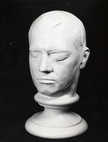
Plaster head cast made of Phineas Gage by Henry Jacob Bigelow at Harvard Medical School in 1850 to substantiate the specifics of Gage’s neurotrauma

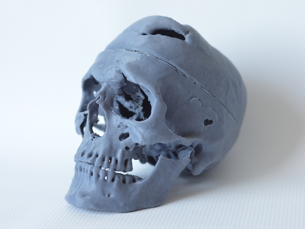
“Beyond the Bone Box” was inspired by Harvard Medical School’s retired bone box program, which enabled medical students to borrow sets of human bones for home study, and developed in partnership with Harvard faculty, curators, archivists, and librarians, this project will develop three circulating resources that contain 3D-printed copies of Warren Anatomical Museum specimens highly contextualized by surrogates of special collections materials. Through this project, the Center seeks to democratize access to unique and sensitive collections through quality fungible surrogates and engender new forms of engagement with Harvard’s special collections across its library system.

The first circulating resource will be a teaching kit built around the case of Phineas Gage, the 19th century railroad foreman whose prefrontal cortex injury has been used to academically and popularly illustrate post-traumatic social disinhibition for the last 150 years.

Project work will begin in September. For the complete list of Lee Innovation Grant award recipients, click here.

Phineas Gage 3D Print!

By , December 5, 2016
Phineas Gage 3D Print, Courtesy of Graham Holt, Laboratories of Cognitive Neuroscience, Boston Children’s Hospital

Phineas Gage 3D Print, Courtesy of Graham Holt, Laboratories of Cognitive Neuroscience, Boston Children’s Hospital

One of the most interesting developments in the renewed teaching capacity and impact of Phineas Gage is the recent establishment of a printable 3D model of well-known patient’s skull. The print file was created by Graham Holt at the Laboratories of Cognitive Neuroscience at Boston Children’s Hospital, and is based on the 2004 thin-slice computed tomography scans of Peter Raitu and Ion-Florin Talos. The file grants a tangible portability to the Gage skull given that the original usually stays safety ensconced in the Warren Museum Exhibit Gallery. Holt’s 3D print had been downloaded 725 times as of October 3rd. The project was featured on the May 5th 3D Printing Today Podcast (segment at 1:02:30). The Warren Anatomical Museum has been using its own version of the Holt print in on-site, hands-on educational programs.

The print file for the Gage skull can be found in the following two places:

The capacity to print a version of Gage’s skull is an exciting addition to the Gage educational experience. More about the original CT scan is discussed in Ratiu, P., Talos, I. F., Haker, S., Lieberman, D., & Everett, P. (2004). “The tale of Phineas Gage, digitally remastered.” Journal of neurotrauma, 21(5), 637-643. More about the Phineas Gage case in general can be found on Malcolm Macmillan’s Phineas Gage Information Page.


Warren Museum Conserves Eustache Belin Phrenology Cast

By , May 25, 2014

Phrenology cast of Eustache Belin, Warren Anatomical Museum, Francis. A. Countway Library [WAM 03235]

Phrenology cast of Eustache Belin, Warren Anatomical Museum, Francis. A. Countway Library [WAM 03235]

The Warren Anatomical Museum recently conserved a phrenology cast of Eustache Belin, formerly of the collection of the Boston Phrenological Society. The Society existed approximately from 1832 to 1842. It possessed a rich cabinet of head, skull and face casts, many of which came from estate of famed phrenologist Johann Gaspar Spurzheim. The Society was formed in Spurzheim’s honor after his death from typhoid in 1832.

The Belin cast was not part of Spurzheim’s collection. The Phrenological Society most likely acquired a copy of a Belin cast from Edinburgh Phrenological Society founder George Combe when his American lecture tour stopped in Boston in 1838. Combe derived his cast from the original cast created by the Paris Phrenological Society. Belin had lived in Paris and died there in 1835.

The phrenologists used the casts of Eustache Belin as evidence of the regions of benevolence and courage. Belin was born into slavery in 1773 in Santo Domingo. During a rebellion on the island in 1791, he was reported to have saved more than 400 people from harm, including the man who held him in slavery, a Monsieur Belin de Villeneuve. Eustache, Monsieur Belin, and a group of refugees escaped Santo Domingo on a boat headed for Baltimore. After this boat was captured by English pirates, Belin led a revolt and overthrew the pirates. When Belin moved to Paris, after obtaining his freedom, he was known to give most of his resources to the disadvantaged.

The Warren Museum’s cast of Belin had suffered a fracture during one of the Boston Phrenological Society collection’s many moves since it was donated to the Medical School by John Collins Warren in 1849. The fracture had split the back of head away from the main cast. Object and art conservator Nina Vinogradskaya attached the broken elements back to the main cast and consolidated and repaired other broken sections of the cast with excellent results.


Warren Museum Conserves New Accessions for “Body of Knowledge” exhibit

By , January 30, 2014
Thomas Dwight lecturing in amphitheater, with Dwight-Emerton skull models, c. 1906., Harvard Medical Library in the Francis A. Countway Library of Medicine

Thomas Dwight, Jr. lecturing using Dwight-Emerton skull models, 1906, Harvard Medical Library in the Francis A. Countway Library of Medicine

In November 2013 Harvard Medical School’s Program in Medical Education generously donated a series of large papier-mâché models designed by Harvard anatomist Thomas Dwight, Jr. and sculptor J. H. Emerton to the Warren Anatomical Museum. Between 1890 and 1895, Emerton made 20 models for Dwight, many of which have survived and are still used in Harvard’s anatomy classrooms. The donation included a 6.5-foot-tall model of a sagittal section of the human skull, a 5-foot-long model of the bones of the foot, various enlarged hand bones and vertebrae – all by Emerton. Also, included in the gift were two papier-mâché Auzoux models, an enlarged ear and a sagittal section of the face with removable layers.

The skull and bones of the foot models are being loaned for the approaching exhibition Body of Knowledge; A History of Anatomy (In Three Parts). The exhibit is a special collaboration of the Center for the History of Medicine, the Collection of Historical Scientific Instruments, Harvard University’s Department of the History of Science, the Harvard Medical School Program in Medical Education, and the Harvard Museums of Science and Culture. The exhibit will open at the Collection of Historical Scientific Instruments on March 6, 2014 and run until December 5, 2014. Approximately 50 anatomical preparations, models, artifacts, books and images  from Center for the History of Medicine collections will be displayed.

Object conservator Nina Vinogradskaya working on Dwight-Emerton skull, Warren Anatomical Museum in the Francis A. Countway Library of Medicine

Object conservator Nina Vinogradskaya working on the Dwight-Emerton skull, Warren Anatomical Museum in the Francis A. Countway Library of Medicine

After 100 years of active teaching, the skull and foot models acquired their fair share of chips, breaks, and abrasions. The Center and the Ackerman Program on Medicine and Culture funded the conservation and partial restoration of the skull and foot. Skilled object conservator Nina Vinogradskaya carefully cleaned the models, consolidated their deteriorating paint layers, repaired breaks in the papier-mâché and plaster, and even restored large sections of loss in the skull’s maxilla and teeth. The models and Nina’s work will be prominently displayed in Body of Knowledge and at the Warren Museum and Countway Library when they return to the Harvard Medical School campus in December 2014.

Harvard Medical School’s Stephanie Dutchen authored an article on the acquisition of the Dwight-Emerton models and their move across the Medical School campus in the school’s news feed.

New artifacts donated to Warren Museum

By , August 8, 2013

Pocket field surgery kit, used in the American Civil War and found on a battlefield in 1862, Warren Anatomical Museum [WAM 21049], Francis A Countway Library of Medicine

Pocket field surgery kit, used in the American Civil War and found on a battlefield in 1862. Warren Anatomical Museum [WAM 21049], Francis A Countway Library of Medicine

The Warren Museum recently acquired two new artifacts.

R. Bryan and Drew Trainor donated a pocket surgery kit to the Museum that had been passed down through their family. The kit was found on an American Civil War battlefield in 1862 by Julius Reed of 1st Regiment of the Heavy Artillery of Connecticut. Reed gave the kit to the donor’s great-great-grandfather.

The surviving instruments in the kit are mostly made by George Tiemann & Co. and were designed for minor surgery. They include an artery forceps, a  tortoise-shell folding probe, a tortoise-shell folding curved bistoury and tenotome, a tortoise-shell folding gum lancet and tenaculum, a tortoise-shell folding curved bistoury and scalpel and a suture needle. The kit also contained a bullet from a Vanderberg Volley Gun.

Harvard Medical School Associate Professor of Medicine Peter Tishler donated a box of Eli Lilly Liver Extract #343, circa 1929, to the Museum’s collection, marking an important moment in the history of hematology as well as the history of university and industry partnerships. In 1926 Harvard physicians George Minot and William Murphy announced that feeding liver to pernicious anemia patients helped restore their health. By 1928 Minot and Murphy had collaborated with Eli Lilly to create and market Liver Extract#343 to treat the disease. Minot and Murphy, along with University of Rochester’s George Whipple, won the 1934 Noble Prize for their pernicious anemia research.

Eli Lilly box containing Liver Extract #343, dated 1929. Warren Anatomical Museum [WAM 21053], Francis A. Countway Library of Medicine

Eli Lilly box containing Liver Extract #343, dated 1929. Warren Anatomical Museum [WAM 21053], Francis A. Countway Library of Medicine

Patients were instructed to take “3 to 6 vials” of Liver Extract #343 a day, depending on the recommendations of their physicians. Each box contained 24 vials of the compound. The donated box most likely came from a former pernicious anemia patient. Of the 16 surviving vials, 15 have been emptied of their contents.

The Warren Museum is grateful to our generous donors whose gifts will benefit of future physicians, historians of science, and the curious public.

Panorama Theme by Themocracy