The BackBlog: Dr. Lloyd T. Brown’s Orthopedic Braces

By , February 25, 2020


In the early stages of the sorting process for this project, while we were still creating guidelines, I found a child’s leg brace in a box of miscellaneous items. The brace started with a hard shoe at the bottom connected to metal splints, which led up to a leather-covered metal band that went around the waist. The tag attached to the brace stated that it was made for a four of five-year-old child to keep the toes from pointing inwards. Later on, I found five other braces from the series in another box. Some braces were for daytime and some were to wear overnight. The smallest ones were for an infant, and the largest was for an eighteen-year-old.

Photo of a child's orthopedic brace with a shoe and attachments at the hip and knee

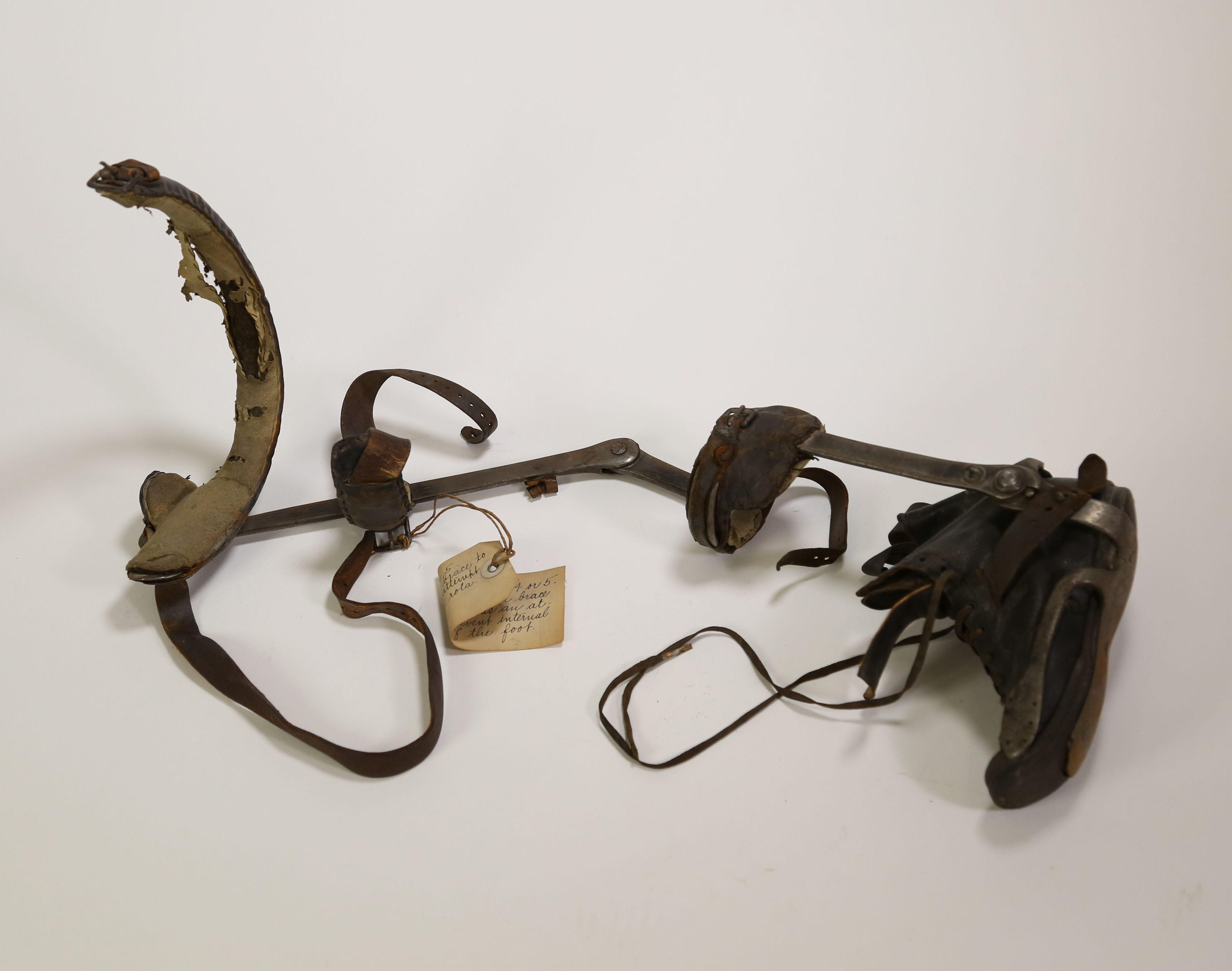
Orthopedic brace for a 4-5 year old. The metal post and waistband help to prevent internal rotation of the foot. Circa 1885. From the Warren Anatomical Museum in the Center for the History of Medicine, Francis A. Countway Library of Medicine (WAM 13255.006A)

Luckily, each brace had a tag with a description, object number, and accession number. This gave me more information to start with than most objects in the backlog did. I was quickly able to find out that Lloyd T. Brown donated this series of braces to the museum in 1943. Based on my initial reading of the accession record, I was under the impression that Brown was the physician who had created the braces for a child. This made sense, as Brown was an orthopedic surgeon. However, the dates didn’t line up, as the earliest braces were made in 1880—the same year that Brown was born. With a bit more research it became clear that Brown was not the physician attached to these objects, but the patient.Lloyd T. Brown was born with a club foot. He was seen by the orthopedic surgeon Edward Hickling Bradford. At the time, Bradford was working at Children’s Hospital and had recently joined the faculty at Harvard Medical School. Bradford was well-known in his field. He co-authored “Orthopedic Surgery” in 1890, which was considered the standard text on orthopedics for many years. He was influential in founding the American Orthopedic Association and was the co-founder of the first school in the United States for children with physical disabilities.

Bradford created the orthopedic braces that I found in the backlog and used them as the primary treatment for Brown during his early childhood. However, this was not enough. Brown received tendon surgery, which helped for some time, and then, at the age of eighteen, a surgery to remove a small amount of bone in his foot. Bradford made one more brace for him post-surgery, and while Brown still had difficulties with his foot throughout his life, this was the last brace that he ever needed.

Brown was so inspired by this experience that he followed in Bradford’s footsteps. Like Bradford, Brown attended Harvard Medical School, where he eventually joined the faculty. He worked at the Massachusetts General Hospital and Children’s Hospital, where he specialized in chronic diseases and orthopedic surgery. During his career, Brown had many patients who were children with disabilities similar to his own. Brown wrote about and discussed his own treatment and felt that it put him in a unique position as a doctor, because he could speak to the results of the treatments later in life.

These orthopedic braces show that medical treatment can have an impact on much more than just physical health. They tell the story of a patient who became a doctor. Lloyd T. Brown chose to donate his childhood braces to the museum, suggesting that he wanted that story to be told. And now that we have rediscovered them, we can tell that story once again.

The BackBlog: The Origins of the Polygraph

By , February 18, 2020

Today, most of us are familiar with the polygraph machine, or, as it is commonly called, the “lie detector”. While some people have encountered the device in real life, most of us have learned about it through pop culture. We’ve seen people strapped to them in every procedural show. If a murder suspect fails a polygraph test or refuses to take one, it’s often considered “proof”—if not admissible evidence—that they are the killer. And of course, they’re a stable of daytime television shows that focus on cheating partners and paternity tests.

Because of this, I was surprised to find a polygraph machine in one of the boxes in our backlog. This machine was given to the museum by George Cheever Shattuck (1879-1972) in 1929. Shattuck was a prominent Boston physician, best known for his work in the field of tropical medicine. Why would a physician have a polygraph machine?

Photograph of a Mackenzie Polygraph. The case containing the object is open to show the parts of the object, which are not assembled.

George C. Shattuck’s Mackenzie Polygraph, 1906-1929. From the Warren Anatomical Museum in the Center for the History of Medicine, Francis A. Countway Library of Medicine (WAM 22202)

Although the polygraph is known colloquially as a lie detector, that isn’t exactly what it does. The machine detects changes in bodily function that indicate stress, which could be a result of lying. This includes functions like breathing, heart rate, and perspiration—all of which are also important baseline health measurements. With this in mind, it makes sense that the original polygraph machine had nothing to do with lies and criminal investigation: it was actually a medical device.

At the turn of the century, Dr. James Mackenzie (1853-1925) developed the first ink-writing polygraph to track a patient’s irregular heartbeat. While simpler than the polygraph that most of us are familiar with today, this device works in much the same way. It features two rubber tambours, one of which was attached to a vein in the neck and the other to the wrist. These tambours would move with the patient’s pulse, and the waves of this movement would be sent down rubber tubing to two recording arms with needles. Then, the needles would record the pulse as a continuous ink-line on paper. The doctor could simply look at the paper to determine the pattern of a patient’s heartbeat.

At the time that he introduced the machine, there wasn’t an effective way for physicians to track the pattern of a patient’s heartbeat. It was replaced by the electrocardiogram machine (or EKG) shortly after, making the Mackenzie polygraph a short but important segment of the history of cardiology. Mackenzie probably never imagined what his machine would eventually become known for, but nevertheless, his legacy continues to this day.

The BackBlog: The Mystery of the Babcock Tester

By , February 11, 2020

I found this device in a box labeled “Misc. Med Equip / Early Autoclave? / + other un-Id’d stuff”. None of the labels on the box seemed quite right for this instrument. It didn’t look exactly like anything I had come across before. Luckily, the name of the object was right on the side, so it didn’t take too long to figure out what it was. The reason I couldn’t place it was that it wasn’t a medical device at all. It was an agricultural device.

Photo of a babcock tester, with a hand-crank and two centrifuge tubes

Hand-cranked Babcock tester, 1890-1940. From the Warren Anatomical Museum in the Center for the History of Medicine, Francis A. Countway Library of Medicine (LEAN0931)

The Babcock Tester was developed in 1890 by Stephen Babcock to detect fat content in milk. The test was simple: place graduated vials of the milk you are testing into a centrifuge like this one and spin them until the milk is separated. Once the milk has separated, you can see the amount of fat that is present in the sample. This tester is a hand-crank model designed for a tabletop, but other iterations included covered centrifuges and table clamps. Sometimes, sulfuric acid was used to remove proteins and other milk components, leaving just the fat.

For the most part, the Babcock test was used by farmers to check the quality of their milk. Sometimes, it was also used to make sure that dairy farmers weren’t diluting their product to stretch the amount of milk they had. The test became incredibly popular and was the primary method for testing milk fat for decades. Not only was it easy and effective, but Babcock refused to patent the device. That made it accessible and affordable as well.

So, if this is an agricultural device, how did the Babcock tester end up in a medical museum? The answer is, we’re not sure. We don’t have any background information on who donated the object or what it was used for.

There are several reasons this Babcock tester could have been collected. It could have been part of a public health initiative regarding nutrition from milk. There might have been a particular physician who was interested in this aspect of nutrition. It’s also possible that someone used this device as a centrifuge for something other than its intended purpose, and it has nothing to do with milk. Hopefully, we will find more information about this device someday, and we will learn how it ended up here. For now, we can only speculate, and the Babcock tester remains one of the mysteries of the backlog.

Staff Finds: Fetal alcohol syndrome education materials

By , February 5, 2020
Covers of pamphlets for expecting parents and "Non-Alcoholic Beverage Recipes" booklet.

Covers of pamphlets for expecting parents and “Non-Alcoholic Beverage Recipes” booklet.

Center staff are currently processing the papers of David Dickinson Potter (1930-2019), who was a founding faculty member in the Department of Neurobiology at Harvard Medical School and co-founder of the Native American High School Summer Program (now named in honor of Potter and his colleague as the Ed Furshpan and David Potter Native American High School Program). The program began as a collaboration between Harvard Medical School and students, teachers, and community members from Native American homelands, and remains a collaborative program today. Potter hosted groups of Hopi, Lakota, Sioux, Wampanoag, Native Hawaiians, and other Native Americans at Harvard each summer, and he worked with the teachers to develop educational curricula that carefully considered the social factors influencing teenagers who lived on Native homelands. The programs often focused on health issues that impacted the students’ communities, such as addiction and fetal alcohol syndrome. Potter’s voluminous files of articles on the effects of alcohol and drugs on the brain attest to his research on neurobiology’s relevance to Native American health. His collection also includes posters, brochures, a word search, a bumper sticker, handwritten notes, and other educational materials about fetal alcohol syndrome from the 1990s.

Pages from "Non-Alcoholic Beverage Recipes" showing recipes for "Berry Blush," "Party Punch," and Water.

Pages from “Non-Alcoholic Beverage Recipes” showing recipes for “Berry Blush,” “Party Punch,” and Water.

This hand-drawn booklet of “Non-Alcoholic Beverage Recipes” was distributed by the Nutrition and Dietetics Training Program in Santa Fe, New Mexico. The booklet contains recipes for non-alcoholic cocktails, such as “Honey Nog” and “Party Punch.” The recipe collection,  informational pamphlets, and bumper sticker, are examples of public health outreach materials aimed at educating Native American communities (or the general population), about the effects of alcohol, particularly on developing fetuses. Most items in Potter’s collection were produced by federal or non-profit organizations, but some items were created by Native American organizations. It was Potter’s close work with Native teachers that led to the creation of in-depth high school education resources that combined neurobiology with the students’ own experiences.

Blue bumper sticker with white text reading "A few drinks can last a lifetime. If you are pregnant...don't drink!"

Bumper sticker created by the National Organization on Fetal Alcohol Syndrome.

In addition to academic work related to neurobiology and health topics, the Native American High School Summer Programs typically included local outings in Boston, and visits to Potter’s homes in Cambridge, Massachusetts for blueberry pancakes and Woods Hole, Massachusetts for fishing. Potter mentored many students from the program long after their visits to Harvard, supporting them as they pursued higher education.

The finding aid for the Potter collection is forthcoming. Some materials may be restricted. Please contact Public Services staff with any questions.

The BackBlog: The Order of the Bifurcated Needle

By , February 4, 2020

As I was going through one of the boxes in our backlog, I found a small blue box. When I opened it up, there was a lapel pin inside. The pin was in the shape of a circle, with one end that went into the back of the pin and one end that was split like a snake’s tongue. There was also a small piece of paper inside with the words “Order of the Bifurcated Needle” in tiny, neat handwriting. I had a feeling that this Order was different than the Order of Saint Michael or the Knights Templar, and I was curious to learn more about it. What I discovered made this tiny object one of the most exciting things that I have found throughout this project.

Photo of a lapel pin in a blue box. The pin is made from a bifurcated needle that has been twisted into a circle.

Lapel pin from the Order of the Bifurcated Needle, 1976. From the Warren Anatomical Museum in the Center for the History of Medicine, Francis A. Countway Library of Medicine (LEAN1158)

In 1966, Donald Ainslie (D.A.) Henderson (1928-2016) became the commanding general of the World Health Organization’s (WHO) smallpox eradication program. Ten years later, he saw that the end of this project was in sight and created something unlike any other honorary organization: The Order of the Bifurcated Needle.

Smallpox was a viral disease that caused a skin rash, resulting in permanent scarring and sometimes loss of vision. The disease had a mortality rate of 30%, with a higher rate amongst infants. Edward Jenner developed a vaccine to protect against smallpox in 1798. The vaccination was given using a bifurcated needle: a short metal rod with a flat, pronged head designed to hold a single dose of the vaccine. As vaccination rates increased amongst developed countries, the disease rate lowered dramatically, but smallpox still proliferated in areas where the vaccine was not easily available. Because of this, WHO determined that smallpox was a good candidate for eradication. They began a campaign for eradication in 1959 but did not see much success until 1966, at which point more funds were allocated to the project and D.A. Henderson became its leader.

At the time that Henderson took on the project, smallpox was endemic in 33 countries. There were an estimated 15 million cases of smallpox every year, with only about 5% reported to health officials. Henderson believed that in order to eradicate the disease, they had to focus on the number of individuals contracting the disease rather than the number of vaccines given. This led him to coin the phrase “Target Zero”, because the goal of the campaign was to see zero cases of smallpox.
After ten years of hard work, Henderson could see that the end was close. He wanted to do something to commemorate the dedication and determination of everyone involved in the eradication process. Together, he and his daughter came up with the idea of the Order of the Bifurcated Needle: an honorary organization whose symbol would be a bifurcated needle twisted into a circle to represent “Target Zero”. Henderson’s daughter, Leigh Henderson, created 700 lapel pins out of the needles, and these pins—along with admittance to the Order—were awarded at the 1976 WHO conference in Geneva. While the order itself may have been a joke, the recognition was sincere. Today, people who were involved in the project still list “Order of the Bifurcated Needle” under their honors.

The last known case of smallpox was reported in 1977, and the WHO declared the disease eradicated in 1980.

Right now, we are not sure who donated this pin to the museum. There was no other information with the box. We are hoping that we can find the answer somewhere in our records.

It’s #ColorOurCollections Week 2020!

By , February 2, 2020

From February 3rd through 7th, cultural institutions from around the world are sharing coloring pages on social media with the hashtag #ColorOurCollections.

Anatomical drawing of arteries in the human neck

Illustration of the external carotid, one of many illustrations in the Center’s 2020 #ColorOurCollections coloring book.

This year, our coloring book includes new and favorite images, anatomical illustrations, therapeutic treatments, and a dancing faun.

We’re sharing our coloring pages here and on Twitter and Instagram (@HarvardHistMed).

Click here to download our entire 2020 coloring book.

Be sure to share your work using the hashtag #ColorOurCollections and we’ll retweet our favorites!

The BackBlog: Elisha Perkins and the Metallic Tractors

By , January 28, 2020

When I found these strange metal objects in one of the first boxes that I opened from the backlog, I knew exactly what they were. These were Perkins tractors: one of the strangest medical fads I had ever heard about.

In 1796, physician Elisha Perkins began selling a new therapeutic device called the Perkins tractor. These tractors were teardrop-shaped metal rods that were about three inches long and flat on the bottom. They were sold in pairs, with one tractor made of iron and the other made of brass. By merely touching these rods to your skin, Perkins claimed, you could cure rheumatism, inflammation, epilepsy, and any number of other medical maladies. The best part of all? There was no need to be a trained doctor in order to use them. All anyone had to do was touch the devices to the afflicted area, and their symptoms would disappear.

Photo of a pair of Perkins Tractors, two teardrop-shaped pieces of metal. One is silver in color and the other is gold.

A pair of Perkins Tractors, circa 1800. From the Warren Anatomical Museum in the Center for the History of Medicine, Francis A. Countway Library of Medicine (LEAN0011)

It might seem obvious to a modern reader that these cure-all devices were too good to be true, but in the late 18th and early 19th century, they were a massive hit. While there were certainly detractors—Perkins was expelled from the Connecticut Medical Society shortly after he began marketing the device—many physicians and noteworthy members of society endorsed his invention. Perkins tractors were sold to physicians and congressmen. Even George Washington bought a pair. The sales were so successful that Perkins quickly branched out to Europe. While Elisha Perkins sold the tractors at home, his son Benjamin Perkins went to England to sell them there, and continued to do so after his father’s death in 1799. The family made quite a bit of money off them. Perkins tractors were sold for $25 a pair–close to a $500 value today.

In the end, however, the skeptics won. Many scientists and doctors felt that the devices were nothing but quackery. Multiple English physicians conducted tests in which some of their patients were treated with genuine Perkins tractors, and some were treated with instruments made of other materials. These physicians all found that all materials were equally effective, so long as the patient believed that they were being treated with genuine Perkins tractors. These were likely some of the earliest experiments into what we know today as the placebo effect: the idea that a person’s body can have a response to a treatment simply because they believe that it will work.

After these experiments were publicized, the Perkins tractors were ridiculed throughout society. Scholars published papers about their ineffectiveness. Poets and cartoonists lampooned the treatment in magazines and newspapers. Eventually, the tractors fell out of fashion, and people stopped using them.

Living in the 21st century, it’s easy to laugh off the Perkins tractors as obvious quackery. However, it’s important to remember that at the time it wasn’t so obvious. Although there were some skeptics, highly educated people fell for these devices. Cases like these remind us that if something seems too good to be true, it very well might be. And 200 years from now, we might be the ones someone’s laughing at.

The BackBlog: Shining a Spotlight on the Warren Anatomical Museum Backlog

By , January 21, 2020

My name is Theodora Burbank, and I’m the Collections Assistant for the Warren Anatomical Museum. In the fall of 2018, the museum created a plan to address our catalog’s backlog of medical instruments. A backlog is something that many museums have, and that can be difficult to get through on top of day-to-day museum work. I have had the pleasure of working on this plan and sorting through the backlog, and now I’m excited to share some of that work with you.

Photo of a row of shelves containing cardboard boxes

Boxes containing medical instruments from the backlog

The Warren Anatomical Museum’s backlog consisted of 104 boxes of historical medical instruments. The majority of these boxes contained objects that were previously housed in an off-site storage facility. Some contained items from a former gallery space. There were also a few boxes of unknown origins. With such a large number of boxes to get through, we realized that we would need to do something different from our usual cataloging process. We looked at all of the data that we would normally take when completing inventory for an object and decided which fields were “essential”—information that a researcher would need to have to find objects or conduct their work. We hoped that by focusing on these essential fields we could get items from the backlog available to researchers as quickly as possible without compromising necessary data.

Before the sorting process began, we didn’t know too much about what we would find. Outside of generalized labels like “Microscopes” and “Plaster Casts”, along with a few peeks into the boxes, the backlog was dark. After 250 hours of sorting through these dark boxes, we learned that the backlog contained almost 4000 individual objects. Some boxes only contained a few objects. Others contained over a hundred. The vast majority of the instruments were from the 19th century, with some as recent as the 1970s and a few dating back to the early 1700s. There were instruments that had been donated by some of the founding members of the Warren’s collection, including Henry Jacob Bigelow and John Collins Warren, as well as many other notable Boston-area physicians. The objects span a wide range of medical fields, with a large concentration of OBGYN and Ear, Nose, and Throat instruments.

In this series of blog posts, I plan to shine a spotlight on some of my favorite finds from the backlog. Some are exciting because of the stories that are connected to them. Others represent major moments in the history of medicine. Still others involved mysteries that needed to be solved. And, of course, some are simply interesting medical devices. Altogether, these objects can teach us not only about the history of medicine but also about the history of the Warren Anatomical Museum and its collections.

I hope that you enjoy this journey through the backlog as much as I have.

Meet the Staff: Stephanie Krauss

By , November 26, 2019

My name is Stephanie Krauss and I have learned so much since starting my job as a Reference Archivist in January 2019. As the reference archivist, I staff the reading room, work with researchers, and assist with exhibitions and events. Since I have started, I have learned a lot about the history of medicine’s intersections with society, specifically with feminism and race. The Center receives a myriad of questions regarding the history of family planning, as we have the papers of John Rock, Abraham Stone, and other physicians involved in the creation of birth control. Each question that comes in gives me an opportunity to learn something new about this important topic!

Additionally, I have really enjoyed working with our rare book collection. Having the opportunity to explore early medical text such as Vesalius and Avicena is such a unique experience. One highlight was when the Center hosted its annual Anatomy Day, gives which the whole Longwood community a chance to explore these early medical texts. I loved having the opportunity to talk to my colleagues and peers about these influential texts and drawings. I am already looking forward to next year’s event.
In my free time, I enjoy running and hiking. I am currently trying to hike all the 4,000 foot mountains in New Hampshire and visit all the United States national parks. I recently started trail running, and I am enjoying exploring trails in the Boston area.

I look forward to continuing to work with the faculty, staff, and students here at the Center for the History of Medicine.

Panorama Theme by Themocracy