Warren Anatomical Museum Drawing in “Visual Science: The Art of Research” exhibition
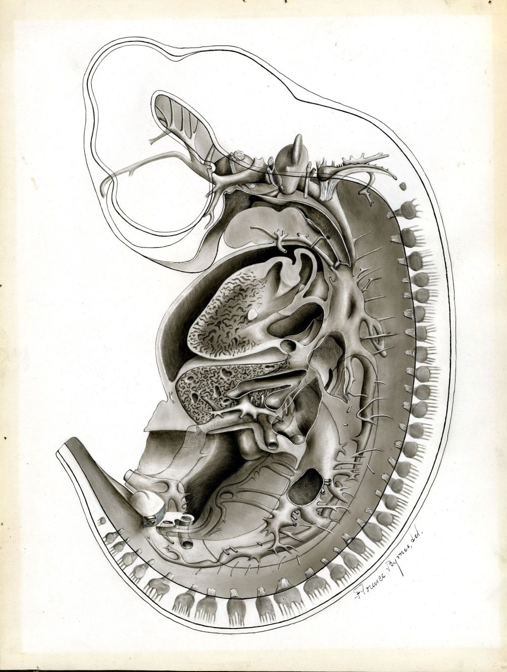
Transverse section of pig embryo at 12 mm, facing, 1903, Warren Anatomical Museum, Center for the History of Medicine, Francis A. Countway Library of Medicine
On September 20, 2019 Harvard’s Collection of Historical Scientific Instruments will be opening an exhibition entitled “Visual Science: The Art of Research.” The exhibition, which features images and objects drawn from a variety of disciplines and time periods that show the importance of visual experiences in science, displays a reproduction of a Warren Anatomical Museum drawing of a pig embryo created in 1903. “Visual Science” is open Sunday – Fridays, 11am–4pm, in the 2nd floor gallery of the Collection of Historical Scientific Instruments.
Harvard Medical School illustrator Florence Byrnes created the original drawing of a transverse section of a pig embryo at 12 mm for Harvard Medical School Professor of Histology and Human Embryology Charles Sedgwick Minot’s 1903 Laboratory Textbook of Embryology. Three other original works by Byrnes of this same pig embryo were also printed in Minot’s textbook.
To make the drawing, Byrnes collaborated with Frederic T. Lewis, then an Instructor in Histology and Embryology. It is a reconstruction derived from hundreds of transverse sections prepared by Lewis. Outlines of individual sections were drawn through a microscope and camera lucida, measured, and compiled into the scale reconstruction by Byrnes. The shading was in part derived from a wax model reconstructed from the embryo sections. Minot believed that reconstructions such as these were highly advantageous in teaching given the very small scale of the original specimens. Despite Minot stating that two of Byrnes’s drawings, including this transverse section of a pig embryo, demonstrated “a special degree of skill and considerable faculty of plastic imagination,” he did not highlight Byrnes as the artist anywhere in the text outside of her signature on the drawings, choosing rather to focus on the histological contribution of Lewis.


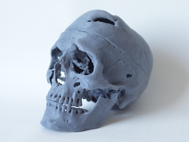
![Phrenology cast of Eustache Belin, Warren Anatomical Museum, Francis. A. Countway Library [WAM 03235]](https://cms.www.countway.harvard.edu/wp/wp-content/uploads/2014/05/03235_v1.jpg)
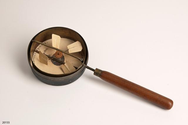
![Pocket field surgery kit, used in the American Civil War and found on a battlefield in 1862, Warren Anatomical Museum [WAM 21049], Francis A Countway Library of Medicine](https://cms.www.countway.harvard.edu/wp/wp-content/uploads/2013/08/21049_v1.jpg)
![Eli Lilly box containing Liver Extract #343, dated 1929. Warren Anatomical Museum [WAM 21053], Francis A. Countway Library of Medicine](https://cms.www.countway.harvard.edu/wp/wp-content/uploads/2013/08/21053_v1.jpg)