Category: Warren Anatomical Museum

Center Receives S.T. Lee Innovation Grant

By , July 10, 2018

The Center for the History of Medicine is pleased to announce that it has received S.T. Lee Innovation Grant funding for its 2018 proposal, “Beyond the Beyond Box.” The application was one of nineteen proposals to bring together Harvard faculty members and library staff; of the nineteen, only six projects were funded. Dominic Hall, Curator, Warren Anatomical Museum, will be spearheading the initiative in partnership with Professor Anne Harrington, Franklin L. Ford Professor of the History of Science.

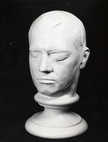
Plaster head cast made of Phineas Gage by Henry Jacob Bigelow at Harvard Medical School in 1850 to substantiate the specifics of Gage’s neurotrauma

“Beyond the Bone Box” was inspired by Harvard Medical School’s retired bone box program, which enabled medical students to borrow sets of human bones for home study, and developed in partnership with Harvard faculty, curators, archivists, and librarians, this project will develop three circulating resources that contain 3D-printed copies of Warren Anatomical Museum specimens highly contextualized by surrogates of special collections materials. Through this project, the Center seeks to democratize access to unique and sensitive collections through quality fungible surrogates and engender new forms of engagement with Harvard’s special collections across its library system.

The first circulating resource will be a teaching kit built around the case of Phineas Gage, the 19th century railroad foreman whose prefrontal cortex injury has been used to academically and popularly illustrate post-traumatic social disinhibition for the last 150 years.

Project work will begin in September. For the complete list of Lee Innovation Grant award recipients, click here.

Phineas Gage 3D Print!

By , December 5, 2016
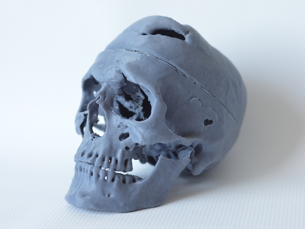
Phineas Gage 3D Print, Courtesy of Graham Holt, Laboratories of Cognitive Neuroscience, Boston Children’s Hospital

Phineas Gage 3D Print, Courtesy of Graham Holt, Laboratories of Cognitive Neuroscience, Boston Children’s Hospital

One of the most interesting developments in the renewed teaching capacity and impact of Phineas Gage is the recent establishment of a printable 3D model of well-known patient’s skull. The print file was created by Graham Holt at the Laboratories of Cognitive Neuroscience at Boston Children’s Hospital, and is based on the 2004 thin-slice computed tomography scans of Peter Raitu and Ion-Florin Talos. The file grants a tangible portability to the Gage skull given that the original usually stays safety ensconced in the Warren Museum Exhibit Gallery. Holt’s 3D print had been downloaded 725 times as of October 3rd. The project was featured on the May 5th 3D Printing Today Podcast (segment at 1:02:30). The Warren Anatomical Museum has been using its own version of the Holt print in on-site, hands-on educational programs.

The print file for the Gage skull can be found in the following two places:

The capacity to print a version of Gage’s skull is an exciting addition to the Gage educational experience. More about the original CT scan is discussed in Ratiu, P., Talos, I. F., Haker, S., Lieberman, D., & Everett, P. (2004). “The tale of Phineas Gage, digitally remastered.” Journal of neurotrauma, 21(5), 637-643. More about the Phineas Gage case in general can be found on Malcolm Macmillan’s Phineas Gage Information Page.


Documenting and preserving the Warren Anatomical Museum’s medical wet specimen collection

By , November 9, 2016


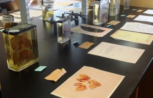
Specimens on display for Anatomy Day, 2016.

Specimens on display for Anatomy Day, 2016.

From roughly the 1840’s through the 1940’s, the Warren Anatomical Museum (WAM) collected and acquired several hundred anatomical wet tissue specimens from medical institutions and from area physicians and academics. While these specimens were originally on display within the museum, the relocation of the WAM to a smaller venue prompted the move of these specimens into storage. In April of 2015, the Museum Collections Technician, Alex Denning, began the task of cataloguing and documenting these specimens. As is the nature with any biological material, the condition of many of the specimens has deteriorated over time, despite preservation efforts. Throughout the history of collecting and saving specimens, chemical preservatives, such as ethanol (or other alcohols and ‘spirits’), formalin and formaldehyde, and various other chemical combinations, have been used to fix (render inert and stable) and preserve anatomical tissues. There is great variability in the current conditions of these specimens and they vary in subject matter from gross anatomical dissection specimens used in teaching, to pathological specimens retained for educational purposes due to their rarity.
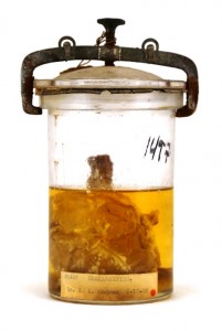
Clavicle with sarcoma.

The task of moving over 800 specimens from museum storage to a laboratory space presented a number of logistical challenges. Due to the nature of the specimens, being housed in a variety of potentially unknown chemical preservatives, relocation of hundreds of medical specimens proves difficult and must be undertaken by special transport. Specimens were properly secured into containers and transported in specialized vehicles to make the journey to the lab. The specimens are now being processed in batches, the first of which contains 300 specimens and is nearing completion.

Processing a historic specimen involves identification, photographing, data collection, cleaning, and repackaging. Each specimen corresponds to a museum number that (hopefully) has documentation on the origins of that particular specimen. Over time, many paper labels have been lost and few specimens have their museum number etched into the glass container or on a tag within the container. This makes identification difficult and many specimens will receive temporary ID numbers until they can be identified through the process of elimination. Within the wealth of information available at WAM and the Center for the History of Medicine (CHoM), often there will be donation or loan histories, and sometimes even patient and surgery information that help to provide context for a particular specimen.

The most important and time-consuming step for each specimen is the data collection. Condition notes are collected, which note the fluid levels and coloration, any deterioration of the specimen, and stability of the container and its seal.  Photo documentation is also a vital step in this process as it records the condition and appearance of a specimen in its found state, which also serves as important data for future processing of specimens. Once documentation is complete, the exterior of the specimen container is cleaned, small cracks are sealed or stabilized, and if possible, a fluid sample is collected for future identification. The specimens are then repacked safely and returned to their storage containers.
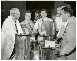
 

Dr. A. T. Hertig, Dept. of Pathology, HMS, using specimens to teach medical students.

Dr. A. T. Hertig, Dept. of Pathology, HMS, using specimens to teach medical students.

As soon as a specimen is fully documented, all data, photographs, and archival information are entered into the WAM database. Efforts are currently underway to make this information available to researchers and the medical community in the future. The project has sought the help of a number of anatomists, pathologists, and medical historians to assess the potential of each specimen for teaching and research purposes. The goal of this project is not just to document and conserve existing specimens, but it is also the hope of the WAM to eventually open specimens up to researchers and scholars upon the project’s completion.
 

Warren Museum device pictured in Harvard Medicine magazine

By , March 1, 2015
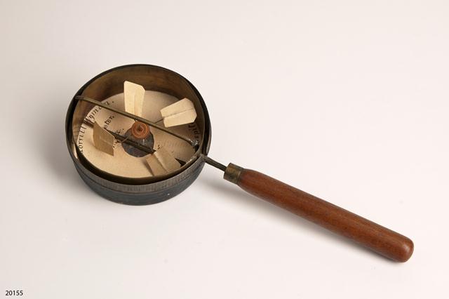
Morrill Wyman "Airmeter", 1867-1903, Warren Anatomical Museum in the Francis A. Countway Library of Medicine (WAM 20155)

Morrill Wyman “Airmeter”, 1867-1903, Warren Anatomical Museum in the Francis A. Countway Library of Medicine (WAM 20155)

An unusual device built by HMS alumnus Morrill Wyman (1812-1903)  is depicted in the  Winter 2015 edition of Harvard Medicine magazine. The device is an airmeter or anemometer, and it was designed by Wyman to register air flow and speed. The device has four paper fan blades set in an open metal circular frame. It has a wooden handle so it could be held up to air vents.

Wyman was a pioneer in recognizing the health consequences of poor ventilation in hospitals and public buildings. In 1846, he won the Boylston Medical prize for an essay on the subject, which he then expanded to a 400-page text entitled A Practical Treatise on Ventilation. Wyman published a paper on the health effects of various outlet cowls for chimneys for the American Academy of Arts and Sciences two years later. As befitted his expertise, he served on an inspection committee of army medical facilities during the American Civil War.

Dr. Wyman had a lifelong dedication to Cambridge Hospital (now Mount Auburn Hospital). He served as its president from 1874 to 1892, and was instrumental in the design of the hospital’s physical plant. His particular interest was a novel and elaborate heating and ventilation system. Venting to the outside was conducted through a main chimney linked to vents placed beneath each bed. It was reported that approximately 2,000 cubic feet of air was drawn from the hospital each hour by the system. Towards the end of his career, Wyman would stop by the hospital on his way home from seeing patients to check on the ventilation system. Accounts had him checking the vents with his airmeter just several weeks before his death at 90.

In addition to the airmeter, the Center for the History of Medicine has an architect’s drawing, circa 1903, of the proposed Harvard Medical School quad, which can be seen in the online version of the Harvard Medicine “Backstory.”

Warren Museum Conserves Eustache Belin Phrenology Cast

By , May 25, 2014

Phrenology cast of Eustache Belin, Warren Anatomical Museum, Francis. A. Countway Library [WAM 03235]

Phrenology cast of Eustache Belin, Warren Anatomical Museum, Francis. A. Countway Library [WAM 03235]

The Warren Anatomical Museum recently conserved a phrenology cast of Eustache Belin, formerly of the collection of the Boston Phrenological Society. The Society existed approximately from 1832 to 1842. It possessed a rich cabinet of head, skull and face casts, many of which came from estate of famed phrenologist Johann Gaspar Spurzheim. The Society was formed in Spurzheim’s honor after his death from typhoid in 1832.

The Belin cast was not part of Spurzheim’s collection. The Phrenological Society most likely acquired a copy of a Belin cast from Edinburgh Phrenological Society founder George Combe when his American lecture tour stopped in Boston in 1838. Combe derived his cast from the original cast created by the Paris Phrenological Society. Belin had lived in Paris and died there in 1835.

The phrenologists used the casts of Eustache Belin as evidence of the regions of benevolence and courage. Belin was born into slavery in 1773 in Santo Domingo. During a rebellion on the island in 1791, he was reported to have saved more than 400 people from harm, including the man who held him in slavery, a Monsieur Belin de Villeneuve. Eustache, Monsieur Belin, and a group of refugees escaped Santo Domingo on a boat headed for Baltimore. After this boat was captured by English pirates, Belin led a revolt and overthrew the pirates. When Belin moved to Paris, after obtaining his freedom, he was known to give most of his resources to the disadvantaged.

The Warren Museum’s cast of Belin had suffered a fracture during one of the Boston Phrenological Society collection’s many moves since it was donated to the Medical School by John Collins Warren in 1849. The fracture had split the back of head away from the main cast. Object and art conservator Nina Vinogradskaya attached the broken elements back to the main cast and consolidated and repaired other broken sections of the cast with excellent results.


Warren Museum Conserves New Accessions for “Body of Knowledge” exhibit

By , January 30, 2014
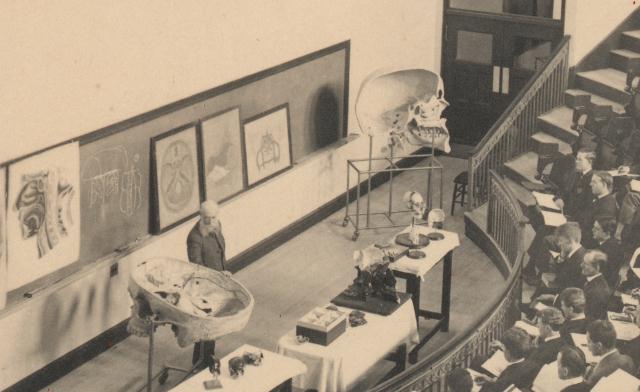
Thomas Dwight lecturing in amphitheater, with Dwight-Emerton skull models, c. 1906., Harvard Medical Library in the Francis A. Countway Library of Medicine

Thomas Dwight, Jr. lecturing using Dwight-Emerton skull models, 1906, Harvard Medical Library in the Francis A. Countway Library of Medicine

In November 2013 Harvard Medical School’s Program in Medical Education generously donated a series of large papier-mâché models designed by Harvard anatomist Thomas Dwight, Jr. and sculptor J. H. Emerton to the Warren Anatomical Museum. Between 1890 and 1895, Emerton made 20 models for Dwight, many of which have survived and are still used in Harvard’s anatomy classrooms. The donation included a 6.5-foot-tall model of a sagittal section of the human skull, a 5-foot-long model of the bones of the foot, various enlarged hand bones and vertebrae – all by Emerton. Also, included in the gift were two papier-mâché Auzoux models, an enlarged ear and a sagittal section of the face with removable layers.

The skull and bones of the foot models are being loaned for the approaching exhibition Body of Knowledge; A History of Anatomy (In Three Parts). The exhibit is a special collaboration of the Center for the History of Medicine, the Collection of Historical Scientific Instruments, Harvard University’s Department of the History of Science, the Harvard Medical School Program in Medical Education, and the Harvard Museums of Science and Culture. The exhibit will open at the Collection of Historical Scientific Instruments on March 6, 2014 and run until December 5, 2014. Approximately 50 anatomical preparations, models, artifacts, books and images  from Center for the History of Medicine collections will be displayed.

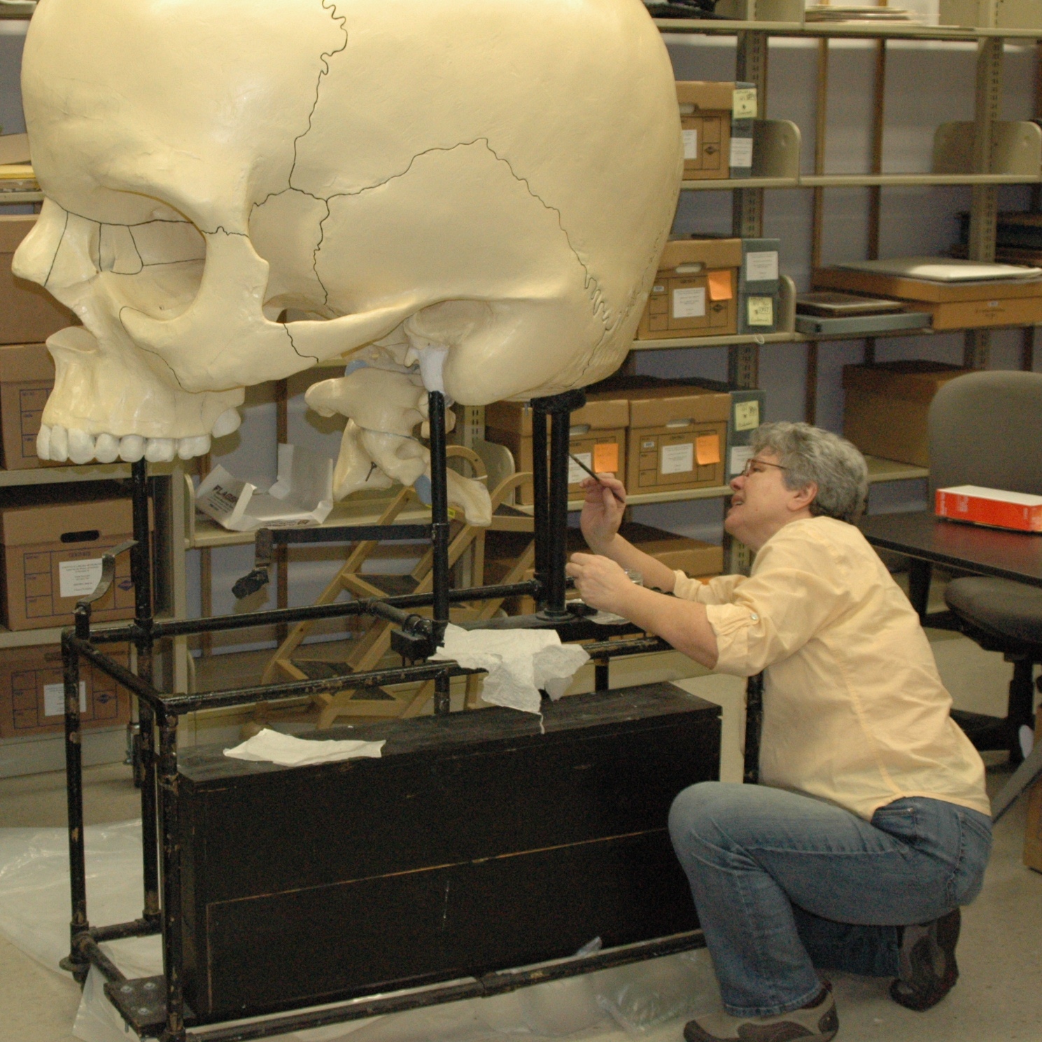
Object conservator Nina Vinogradskaya working on Dwight-Emerton skull, Warren Anatomical Museum in the Francis A. Countway Library of Medicine

Object conservator Nina Vinogradskaya working on the Dwight-Emerton skull, Warren Anatomical Museum in the Francis A. Countway Library of Medicine

After 100 years of active teaching, the skull and foot models acquired their fair share of chips, breaks, and abrasions. The Center and the Ackerman Program on Medicine and Culture funded the conservation and partial restoration of the skull and foot. Skilled object conservator Nina Vinogradskaya carefully cleaned the models, consolidated their deteriorating paint layers, repaired breaks in the papier-mâché and plaster, and even restored large sections of loss in the skull’s maxilla and teeth. The models and Nina’s work will be prominently displayed in Body of Knowledge and at the Warren Museum and Countway Library when they return to the Harvard Medical School campus in December 2014.

Harvard Medical School’s Stephanie Dutchen authored an article on the acquisition of the Dwight-Emerton models and their move across the Medical School campus in the school’s news feed.

New artifacts donated to Warren Museum

By , August 8, 2013

Pocket field surgery kit, used in the American Civil War and found on a battlefield in 1862, Warren Anatomical Museum [WAM 21049], Francis A Countway Library of Medicine

Pocket field surgery kit, used in the American Civil War and found on a battlefield in 1862. Warren Anatomical Museum [WAM 21049], Francis A Countway Library of Medicine

The Warren Museum recently acquired two new artifacts.

R. Bryan and Drew Trainor donated a pocket surgery kit to the Museum that had been passed down through their family. The kit was found on an American Civil War battlefield in 1862 by Julius Reed of 1st Regiment of the Heavy Artillery of Connecticut. Reed gave the kit to the donor’s great-great-grandfather.

The surviving instruments in the kit are mostly made by George Tiemann & Co. and were designed for minor surgery. They include an artery forceps, a  tortoise-shell folding probe, a tortoise-shell folding curved bistoury and tenotome, a tortoise-shell folding gum lancet and tenaculum, a tortoise-shell folding curved bistoury and scalpel and a suture needle. The kit also contained a bullet from a Vanderberg Volley Gun.

Harvard Medical School Associate Professor of Medicine Peter Tishler donated a box of Eli Lilly Liver Extract #343, circa 1929, to the Museum’s collection, marking an important moment in the history of hematology as well as the history of university and industry partnerships. In 1926 Harvard physicians George Minot and William Murphy announced that feeding liver to pernicious anemia patients helped restore their health. By 1928 Minot and Murphy had collaborated with Eli Lilly to create and market Liver Extract#343 to treat the disease. Minot and Murphy, along with University of Rochester’s George Whipple, won the 1934 Noble Prize for their pernicious anemia research.

Eli Lilly box containing Liver Extract #343, dated 1929. Warren Anatomical Museum [WAM 21053], Francis A. Countway Library of Medicine

Eli Lilly box containing Liver Extract #343, dated 1929. Warren Anatomical Museum [WAM 21053], Francis A. Countway Library of Medicine

Patients were instructed to take “3 to 6 vials” of Liver Extract #343 a day, depending on the recommendations of their physicians. Each box contained 24 vials of the compound. The donated box most likely came from a former pernicious anemia patient. Of the 16 surviving vials, 15 have been emptied of their contents.

The Warren Museum is grateful to our generous donors whose gifts will benefit of future physicians, historians of science, and the curious public.

Warren Museum medical case featured in Journal of Bone and Joint Surgery

By , September 17, 2012

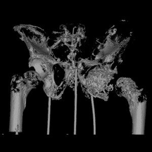
Still of Charles Lowell CT scan, 2009, WAM 07877, Warren Anatomical Museum, Francis A. Countway Library of Medicine

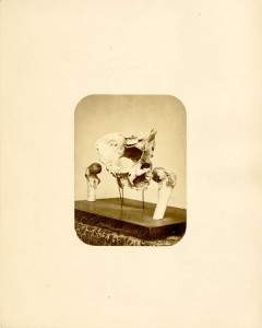
In the September 5th edition of The Journal of Bone & Joint Surgery, William H. and Johanna A. Harris Professor of Orthopedic Surgery James H. Herndon, M.D. published an orthopedic and historical analysis of one of the Warren Museum’s most compelling medical cases, the sacrum, pelvis and femur upper extremities of an early 19th century Maine resident named Charles Lowell. Lowell is recognized as MGH’s first orthopedic case and his accident and subsequent malpractice claims are cited as one of earliest, well-published medical trials in the Unites States.

On September 7, 1821 Lowell was thrown from his horse in Lubec, Maine, dislocating his left hip. His two physicians, John Faxon and Micajah Hawkes, believed they successful reduced the hip but when Hawkes visited Lowell 4-6 weeks later, he found that the injury persisted. Lowell traveled to the newly found Massachusetts General Hospital and its chief surgeon [and Warren Museum founder] John Collins Warren for relief. After much effort, Warren was unable to reduce the injury. Lowell left Boston vowing to sue Faxon and Hawkes for failing to repair his hip.

Lowell vs. Faxon and Hawkes went through three trials. In June 1822 a jury found for Lowell. An appeal in September ended in a hung jury. The final trial in 1824 was the most involved, with many physicians, including John Collins Warren testifying. Warren later published his remarks in an explanatory pamphlet. The case ended in the physicians’ favor. As the specifics of his injury proved the crux of the trial, Lowell was determined to have a postmortem done and upon his 1858 death Jonathan Mason Warren [John Collins Warren’s son] sent a colleague to perform the autopsy, bringing the hip back to Boston with family consent for further examination. This was the last orthopedic analysis done of Lowell’s injury – until Herndon’s 2010 study.

Lowell’s hip preparation was transferred to the Warren Museum by J. Collins Warren [John Collins Warren’s grandson] and accessioned into the collection circa 1885. In 2010 James Herndon, with

Historical photograph of Lowell hip preparation, 1858-1915, WAM 07877, Warren Anatomical Museum, Francis A. Countway Library of Medicine

permission from the Museum, took radiographs and a CT scan of Lowell’s hip. His findings, entitled  An Orthopaedic Case Contributed Substantially to the First Malpractice Crisis in the United States in the Nineteenth Century, have been published in Volume 94-A, Number 17 of the Journal of Bone and Joint Surgery. Dr. Herndon generously donated copies of the scans and radiographs to the Warren Museum to benefit future researchers.

The Charles Lowell pelvis and femur preparation can be found exhibited in the Warren Museum Exhibition Gallery on the 5th floor of the Countway Library of Medicine on the Harvard Medical School campus. More information on visiting the Gallery can be found here.

Warren Museum bladder calculus featured on WBUR’s Radio Boston

By , February 4, 2011

Bladder Calculus, 1809, 04809, Warren Anatomical Museum, Francis A. Countway Library of Medicine

Warren Museum bladder calculus 04809 was recently featured on WBUR’s Radio Boston.

The program toured Harvard University’s new exhibition Tangible Things and interviewed the exhibit curators Laurel Thatcher Ulrich, the 300th Anniversary University Professor in Department of History and Ivan Gaskell, the Margaret S. Winthrop Curator and Senior Lecturer on History.

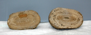
The large stone will be on display at the Harvard Museum of Natural History from January 24 through May 29, 2011. The stone was removed post mortem by a Salem, MA surgeon from a man’s bladder in 1809. According to a historical analysis, it is composed of ammonium urate and uric acid.

The entire Radio Boston story can be found on the WBUR website.

Seventeen artifacts and specimens from the Center for the History of Medicine are on display in Tangible Things. More information on the exhibition can be found in the Center for the History of Medicine News.

Panorama Theme by Themocracy