Category: Warren Anatomical Museum

Warren Anatomical Museum Drawing in “Visual Science: The Art of Research” exhibition

By , September 19, 2019

Transverse section of pig embryo at 12 mm, facing, 1903, Warren Anatomical Museum, Center for the History of Medicine, Francis A. Countway Library of Medicine

On September 20, 2019 Harvard’s Collection of Historical Scientific Instruments will be opening an exhibition entitled “Visual Science: The Art of Research.” The exhibition, which features images and objects drawn from a variety of disciplines and time periods that show the importance of visual experiences in science, displays a reproduction of a Warren Anatomical Museum drawing of a pig embryo created in 1903. “Visual Science” is open Sunday – Fridays, 11am–4pm, in the 2nd floor gallery of the Collection of Historical Scientific Instruments.

 Harvard Medical School illustrator Florence Byrnes created the original drawing of a transverse section of a pig embryo at 12 mm for Harvard Medical School Professor of Histology and Human Embryology Charles Sedgwick Minot’s 1903 Laboratory Textbook of Embryology. Three other original works by Byrnes of this same pig embryo were also printed in Minot’s textbook.

To make the drawing, Byrnes collaborated with Frederic T. Lewis, then an Instructor in Histology and Embryology. It is a reconstruction derived from hundreds of transverse sections prepared by Lewis. Outlines of individual sections were drawn through a microscope and camera lucida, measured, and compiled into the scale reconstruction by Byrnes. The shading was in part derived from a wax model reconstructed from the embryo sections. Minot believed that reconstructions such as these were highly advantageous in teaching given the very small scale of the original specimens. Despite Minot stating that two of Byrnes’s drawings, including this transverse section of a pig embryo, demonstrated “a special degree of skill and considerable faculty of plastic imagination,” he did not highlight Byrnes as the artist anywhere in the text outside of her signature on the drawings, choosing rather to focus on the histological contribution of Lewis.

Warren Anatomical Museum Exhibition Gallery Now Closed Until Spring 2021

By , August 21, 2019

Life cast and skull of Phineas Gage, Warren Anatomical Museum, Center for the History of Medicine, Countway Library

As of Wednesday, 8/21/2019, the Warren Anatomical Museum exhibition gallery will be closed until Spring 2021 to prepare for its redesign as part of the larger renovation of the Countway Library of Medicine. Throughout August, the exhibits will be taken down both for their protection during the upcoming construction and to allow for the curation of the next iteration of the Warren Anatomical Museum exhibition gallery.

Keep an eye out on the Center for the History Medicine news feed and the Countway Library website for updates on the renovation and ways you can give input on the next Warren Anatomical Museum gallery.

Warren Anatomical Museum Gallery Temporarily Closing Until Spring 2021

By , August 14, 2019

Skull of Phineas Gage, Warren Anatomical Museum, Center for the History of Medicine, Francis A. Countway Library of Medicine

This fall, the Francis A. Countway Library of Medicine will begin a multi-floor renovation requiring the temporary deinstallation of the Center for the History of Medicine’s Warren Anatomical Museum Gallery on the fifth floor. Throughout August, all of the artwork, artifacts, and specimens in the building are being taken down for their protection during construction. Plans are now underway to design and imagine the Gallery for its re-installation, including bringing new selections from the Museum’s rich holdings to the public Spring 2021.

The Warren Anatomical Museum will continue to offer educational sessions throughout the renovation and host a number of open houses during the fall and spring academic semesters. Please visit our event calendar this September for these limited-attendance programs. Also look for new content about Museum holdings on our new Museum landing page.

The last day to visit the Gallery and the skull of Phineas Gage is Tuesday, August 20, 2019.


Warren Museum’s Mystery Box Reveals a “Twilight” Story

By , July 25, 2019

Doctor’s Birthing Kit, circa 1910

Anesthesia history artifacts collected by Bert B. Hershenson, MD

Anesthesia history artifacts collected by Bert B. Hershenson, MD

This mysterious metal box filled with labeled glass bottles and anesthesia paraphernalia was one of the anesthesia history artifacts collected by Bert B. Hershenson, MD, Director of Anesthesia (1942–1956) at the Boston Lying-in Hospital (a Brigham and Women’s parent hospital). It was donated by Mrs. Hershenson to Harvard Medical School’s Warren Anatomical Museum in 1972 with no identifying information other than that it once belonged to a Viennese doctor “two generations ago.” A recent provenance investigation of the box and the objects inside, done here at the Center for the History of Medicine, indicated that the original owner was probably a turn-of-the-century obstetrician who may have been a practitioner of Dämmerschlaf or “Twilight Sleep.”

Picture of the March 7, 1915 Boston Sundat Post newspaper article, "Scores of Twilight Sleep Babies in Hub"

Boston Sunday Post, March 7, 1915. “Scores of Twilight Sleep Babies in Hub”

Twilight Sleep was introduced in Germany at the beginning of the 20th century. A combination of morphine, to mitigate pain, and scopolamine to cause amnesia, was given by injection to women in labor. Its effectiveness in preventing pain was minimal. Its true effectiveness was in causing many women to forget the pain and the subsequent extreme, sometimes violent, behavior the drug combination often caused. In 1914, reports of “pain free” deliveries in Europe gave rise in the U.S. to the National Twilight Sleep Association, which successfully campaigned for the widespread adoption of the technique. However, in 1915 Mrs. Francis X. Carmody, a leader of the organization, died in childbirth. Although probably unrelated to the drugs, news of her death and subsequent safety concerns caused a fall from favor of Twilight Sleep in America and the end of the Association. Newer variations on the technique did continue through the 1960s until the advent of the natural childbirth movement.

Object list:

Metal box (for easy sterilization) from medical supply house Medicinisches Waarenhaus: Berlin

Esmarch type inhaler (style introduced in 1877). The wire mask covered by a cloth kept chloroform from touching the patient’s face.

Chloroform, a surgical anesthetic.

Erogotin, used to treat excessive bleeding and to speed up labor.

Camphor, traditionally used as a topical analgesic, or to control nausea.

Morphium, for pain relief.

Unidentified bottle, with the handwritten word “injection’ in German.

Dr. Vomel brand catgut, probably used for tying off the umbilical cord.

Warren Anatomical Museum Collection, Center for the History of Medicine
in the Francis A. Countway Library of Medicine

Event Video Now Available: Human Tissue Ethics in Anatomy, Past and Present

By , July 25, 2019

The Center for the History of Medicine is pleased to announce that the recording of the April 4, 2019 symposium, Human Tissue Ethics in Anatomy, Past and Present: From Bodies to Tissues to Data, is now online. The symposium, which was co-sponsored by Harvard University’s Ackerman Program on Medicine and Culture, the Center for the History of Medicine in the Francis A. Countway Library, Harvard Medical School’s Department of Global Health and Social Medicine, and the Department of Pediatrics, Boston Children’s Hospital, explored transparent and ethical anatomical body and tissue procurement as a cornerstone of medical ethics in research and education. Watch the Tissue Ethics symposium.


Symposium Contents

Panel 1: Human Tissue Ethics in Historical Contexts of Anatomy
Scott H. Podolsky, Harvard Medical School, Chair

  • Dominic W. Hall, Harvard Medical School: The Second Life of Specimens: Scientific and Historical Research in the Warren Anatomical Museum
  • Sabine Hildebrandt, Harvard Medical School/Boston Children’s Hospital: Dealing with Legacies of Nazi Anatomy: the ‘Vienna Protocol’
  • Tinne Claes, Katholieke Universiteit: Why Is It So Difficult to Throw Away Fetuses? Anatomical Collections and the Meanings of Disposal
Speaker Sabine Hildebrandt presenting her talk, Dealing with Legacies of Nazi Anatomy: the ‘Vienna Protocol’

Speaker Sabine Hildebrandt giving her talk, Dealing with Legacies of Nazi Anatomy: the ‘Vienna Protocol’


Panel 2: Human Tissue Ethics in Current Anatomical Education and Research
Dan Wikler, Harvard T.H. Chan School of Public Health, Chair

  • Thomas Champney, University of Miami: The Business of Bodies: Human Tissue Ethics and Commercialization
  • Michel Anteby, Boston University: Nested Moralities: From National to Intimate Cadaver Trades


Thomas Champney giving his talk, The Business of Bodies: Human Tissue Ethics and Commercialization

Thomas Champney giving his talk, The Business of Bodies: Human Tissue Ethics and Commercialization


Panel 3: Human Tissue Ethics from Physical Specimens to Data
David S. Jones, Harvard University, Chair

  • Maria Olejaz Tellerup, University of Copenhagen: The Anatomy of Bioavailability: Exploring Body Donation in Denmark Then, Now and in the Future
  • Jon Cornwall, University of Otago: The Impact of Digital Technology on Body Donation
The Anatomy of Bioavailability: Exploring Body Donation in Denmark Then, Now and in the Future

Maria Olejaz Tellerup giving her talk, The Anatomy of Bioavailability: Exploring Body Donation in Denmark Then, Now and in the Future


Register now for Normalizing Sex Research and Education in America: Robert Latou Dickinson in Perspective (5/23)

By , May 14, 2019

Upcoming event!

Normalizing Sex Research and Education in America: Robert Latou Dickinson in Perspective

Registration is required

Physician Robert Latou Dickinson (1861–1950) resists categorization. He was a long-time obstetrician and gynecologist; a research scientist invested in sexual health who influenced Alfred Kinsey and notions of sexuality; a birth control and reproductive sterilization advocate; an anatomist who authored an influential atlas of reproductive anatomy/ an artist who illustrated his own scientific texts; and a public health educator whose popular sculptures and models changed the way the public visualized the birth process. “Normalizing Sex Research and Education in America: Robert Latou Dickinson in Perspective” will explore different aspects of Dickinson’s long career, addressing his work in reproductive health and family planning, his time spent as a sex educator and artist at the New York Academy of Medicine, his Birth Series models created for the 1939 World’s Fair, and his depictions of human anatomy and concepts of normalization through his models Norma and Normman. Dickinson’s legacy is still with us today, and his personal papers and models remain some of the Center for the History of Medicine’s most-used collections. With the help of these four scholars, we hope to better understand the impact and legacy that Robert Latou Dickinson continues to exert on our current health science and clinical care community.

An exhibition also entitled Normalizing Sex Research and Education in America: Robert Latou Dickinson in Perspective will be on display for the event (L1 of the Countway Library of Medicine).

Robert Latou Dickinson

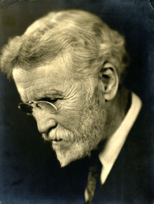
Robert Latou Dickinson (1861–1950), 1913. From the Robert Latou Dickinson papers, 1881-1972 (inclusive), 1926-1951 (bulk) (B MS c72) in the Boston Medical Library, Francis A. Countway Library of Medicine


Speakers

Sarah B. Rodriguez, Ph.D., Senior Lecturer, Global Health Studies, Weinberg College of Arts & Sciences; Lecturer, Medical Education, Feinberg School of Medicine; Faculty, Medical Humanities & Bioethics Graduate Program, all at Northwestern University, Robert Latou Dickinson: Pioneering Researcher.

A founding father of sex research in the United States, a prominent physician who used his position to advocate for access to birth control, and a distinguished clinician: Robert Latou Dickinson, with his deep interest in women’s health, took on all of these roles. In this presentation, Rodriguez will discuss these three roles – sex researcher, birth control advocate, and clinician – of this historically understudied physician, focusing on his pioneering research regarding female sexuality.

Anne Garner, MLS, Curator, Rare Books and Manuscripts Library, The New York Academy of Medicine Library, From the stacks to the studio:  Robert Latou Dickinson’s Academy of Medicine.

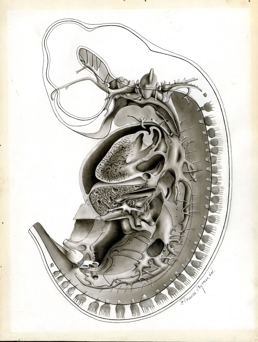
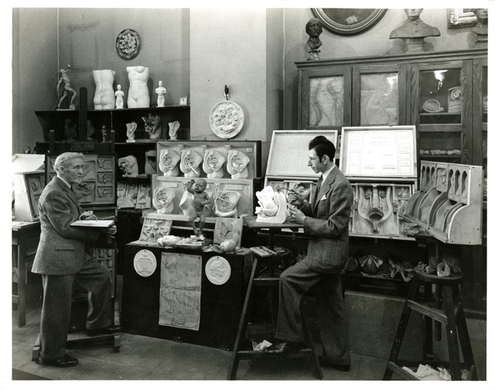
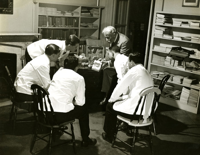
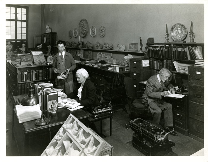
Robert Latou Dickinson’s relationship with the New York Academy of Medicine was a critical part of both his professional and creative identity. In 1891, Dickinson became a Fellow of the organization and served on numerous committees, including as Chairman of the Academy’s art committee from 1935-1940.  At the Academy Dickinson was given a dedicated studio space, where he worked on the Birth Series and other three-dimensional anatomical models. While de facto artist-in-residence, Dickinson also engaged Alfred Kinsey to lobby the Academy to open a sex education library. This talk will explore Dickinson’s role as influencer and occasional disrupter within the Academy, as he advocated for sex education and for greater access to medical information for public audiences.

Rosemarie Holz, Ph.D., Associate Professor of Practice, Associate Director, Women’s and Gender Studies, University of Nebraska-Lincoln, “So that which has been lost is now found!” Exploring the magic of the 1939 Dickinson-Belskie Birth Series Sculptures.”

In this presentation Holz will discuss the creation and dissemination of the hugely influential yet surprisingly overlooked 1939 Dickinson-Belskie Birth Series sculptures, which illustrate the process of human development from fertilization through delivery. First displayed at the 1939-1940 World’s Fair in New York City, they were reproduced in a variety of forms and sent out across the United States and overseas, giving rise to modern views of pregnancy decades before Lennart Nilsson’s much-heralded in utero photographs in Life magazine in the 1960s. Despite their enormous popularity, by the 1970s and ‘80s the Birth Series began to disappear from public knowledge, eclipsed by new technologies, such as ultrasound, that offered modern ways to view in utero development. Holz will conclude her presentation by describing the Birth Series’ surprising re-birth since their 2014 recovery from the dusty storage collection of the University of Nebraska State Museum, a re-birth that is prompting renewed fascination with these evocative forms and new conversations.

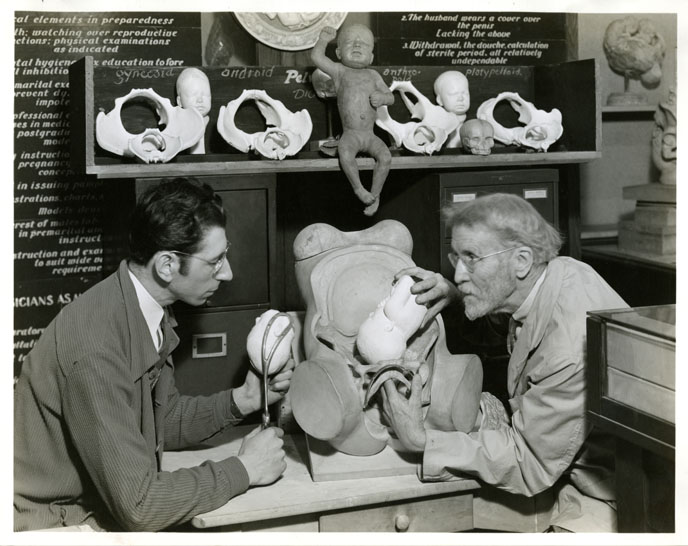
Anna Creadick, Ph.D., Professor of English and American Studies, Hobart and William Smith Colleges, Model bodies, normal curves: Norm and Norma in Postwar America.

In the early 1940s, with their Birth Series models completed, sexologist Robert L. Dickinson and his sculptor-collaborator Abram Belskie created two anthropometric sculptures representing the “average” American male and female bodies. Dickinson named them “Normman and Norma.” Dickinson’s effort to model the “normal” body was indicative of a broader obsession taking hold in midcentury America, as doctors, psychiatrists, physical anthropologists, and scientists began to isolate the normal as a subject, to try and define normality with increasing precision.  In 1945, Dickinson donated the “Norm and Norma” statues along with his Birth Series and other medical models to the Cleveland Health Museum, where they might have just gathered dust. But instead, Norm and Norma began a decade-long tour of the postwar public sphere, appearing in newspaper articles, popular science magazines, television shows, and even a look-alike contest. This presentation tells the story of the wartime production and postwar reception of these models, whose “normal” curves helped to promote a powerful organizing category of postwar culture

Collection link: https://collections.countway.harvard.edu/onview/collections/show/132


Sign up for a Spring Tour!

By , April 5, 2019

Join us for one of the many tours the Center is offering this spring. Tours are free and open to the public, but registration is required.

Leder, Philip, “Codons notebooks,” OnView: Digital Collections & Exhibits, accessed April 5, 2019, http://collections.countway. harvard.edu/onview/items/show/13021.

Highlights from Center for the History of Medicine Collections
The Center for the History of Medicine in the Countway Library is a hub of activity for the history of medicine and society in Boston’s Longwood Medical Area. Located on L2 of the Countway Library, the Center is home to over 2,790 manuscript collections and institutional records series, 1,256 gigabytes of migrated born digital records, 177,676 rare books and journals, and 15,768 Warren Anatomical Museum objects (including artifacts, anatomical, osteological and fluid preparations). Join Jessica Murphy, the Center’s Public Services Librarian, for an introduction to selections that are not only unique to Harvard, but inform and deepen our understanding of contemporary medicine. Register.

Public Health and Harvard: Selections from Center for the History of Medicine Collections
Join Heather Mumford, Archivist for the Harvard T.H. Chan School of Public Health, for an introduction to manuscript and archival collections that illustrate the rich and innovative history of the Chan School of Public Health. Selections from the Center’s historical collections will be on display as part of the tour. Register.

Diversity, Inclusion, and the Medical School Archives
Join Joan Ilacqua, Center for the History of Medicine’s Archivist for Diversity and Inclusion, for a history of those underrepresented in medicine at Harvard through the manuscript and archival collections held by the Countway Library. The tour will include a discussion of current collecting initiatives and a display of selections from the Center’s rich historical collections. Register.

Curator’s Tour of the Warren Anatomical Museum
The Warren Anatomical Museum is one of the last surviving anatomy and pathology museum collections in the United States. In 1847, Harvard anatomist and surgeon John Collins Warren founded the Museum to preserve and classify specimens and models needed for teaching. Until 1999, the Museum was in the Department of Anatomy and Cell Biology. It is now an integral part of the Countway Library’s Center for the History of Medicine.

Join Museum Curator Dominic Hall for a guided tour of the Museum holdings on exhibit in the Countway Library, including the skull, life cast, and tamping iron of Phineas Gage. Register.

Register Now! Human Tissue Ethics in Anatomy, Past and Present: From Bodies to Tissues to Data

By , March 6, 2019

9:00am-3:00pm, Thursday, April 4, 2019
Waterhouse Room, Gordon Hall, Harvard Medical School Campus

Co-sponsored by the Ackerman Program on Medicine and Culture, Harvard University; the Center for the History of Medicine in the Francis A. Countway Library; the Department of Global Health and Social Medicine, Harvard Medical School; and the Department of Pediatrics, Boston Children’s Hospital

NIH Technicians (ID 2263)

Technicians examining plates and tissue culture flasks at a laminar flow hood, 1986. Courtesy National Cancer Institute, National Institutes of Health (ID 2263).

Anatomy as a science and as an educational discipline in the medical curriculum is forever in transition. One of the greatest areas of change in recent decades has been the systematic evaluation of ethical questions in anatomy. At the center of these deliberations is the status of the dead human body, which is no longer only seen as a mere “object” or “material” of research or as an educational “tool.” Rather, it is described as a body that still has connections with the person who once inhabited it, thus becoming part of a social network of knowledge gain and requiring respectful treatment.

This change of perspective will be explored in the symposium, “Human Tissue Ethics in Anatomy, Past and Present: From Bodies to Tissues to Data.” An international group of scholars will discuss the ethical aspects of existing questions, explore the relevance of non-profit and for-profit body donation, and examine newly emerging technologies in anatomy that may need innovative ethical approaches. The aim of this symposium is to present evidence for the insight that transparent and ethical anatomical body and tissue procurement is indeed at the core of medical ethics in research and education.

Registration is required. Register here.


PROGRAM

9:00-10:30am
Panel 1: Human Tissue Ethics in Historical Contexts of Anatomy:
Scott H. Podolsky, Harvard Medical School, Chair

  • Dominic Hall, Harvard Medical School: The Second Life of Specimens: Scientific and Historical Research in the Warren Anatomical Museum
  • Sabine Hildebrandt, Harvard Medical School/Boston Children’s Hospital: Dealing with Legacies of Nazi Anatomy: the ‘Vienna Protocol’
  • Tinne Claes, Katholieke Universiteit: Why Is It So Difficult to Throw Away Fetuses? Anatomical Collections and the Meanings of Disposal

10:30-11:00am
Break

11:00-12:30pm
Panel 2: Human Tissue Ethics in Current Anatomical Education and Research:
Dan Wikler, Harvard T.H. Chan School of Public Health, Chair

  • Thomas Champney, University of Miami: The Business of Bodies: Human Tissue Ethics and Commercialization
  • Michel Anteby, Boston University: Nested Moralities: From National to Intimate Cadaver Trades
  • Glenn Cohen, Harvard Law School/Petrie-Flom Center: The Law and Ethics of Tissue Ownership

12:30-1:30pm
Lunch (provided)

1:30-3:00pm
Panel 3: Human Tissue Ethics from Physical Specimens to Data:
David S. Jones, Harvard University, Chair

  • Maria Olejaz Tellerup, University of Copenhagen: The Anatomy of Bioavailability: Exploring Body Donation in Denmark Then, Now and in the Future
  • Jon Cornwall, University of Otago: The Impact of Digital Technology on Body Donation

Image: Technicians examining plates and tissue culture flasks at a laminar flow hood, 1986. National Cancer Institute, National Institutes of Health (ID 2263, 1986).

Related LibGuide: Searching the Warren Anatomical Museum collection by Dominic Hall


Staff Finds: Dickinson-Belskie

By , February 19, 2019
0004074_ref

Robert Latou Dickinson

While processing the Robert Latou Dickinson papers, Center staff came across photographs of Robert Latou Dicksinon and artist Abram Belskie in their studio at the New York Academy of Medicine.

After retirement from medical practice, Dickinson collaborated with Belskie at the New York Academy of Medicine to create molds and models of human reproductive anatomy for display and as teaching aids, some of which were exhibited at World’s Fair in 1939. These models were later featured in the teaching book Birth Atlas. The sculptures Norma and Normman were a product of their collaboration as well. In 1945, Dickinson transferred his reproductive anatomy model collection to the Cleveland Health Museum. In 2007 the Health Museum was absorbed into the Cleveland Museum of Natural History and at that time transferred the Dickinson-Belskie collection to the Warren Anatomical Museum.

Below are photographs of Dickinson and Belskie in studio with their teaching models, from the Dickinson papers. The photographs were originally produced for Look Magazine; the Library of Congress now holds the copyright for the images. (LOOK Magazine Photograph Collection, Library of Congress, Prints & Photographs Division)

The Dickinson-Belskie Collection in the Warren Anatomical Museum (object numbers 20500-20778) is available for research. Advance notice is required.

The finding aid for the Dickinson papers can be found here.

For information regarding access to these collections, please contact the Public Services staff.

Panorama Theme by Themocracy